Clear Sky Science · en
Left atrial strain response to acute preload reduction in healthy dogs using a translational blood donation model
Why this study matters for pets and people
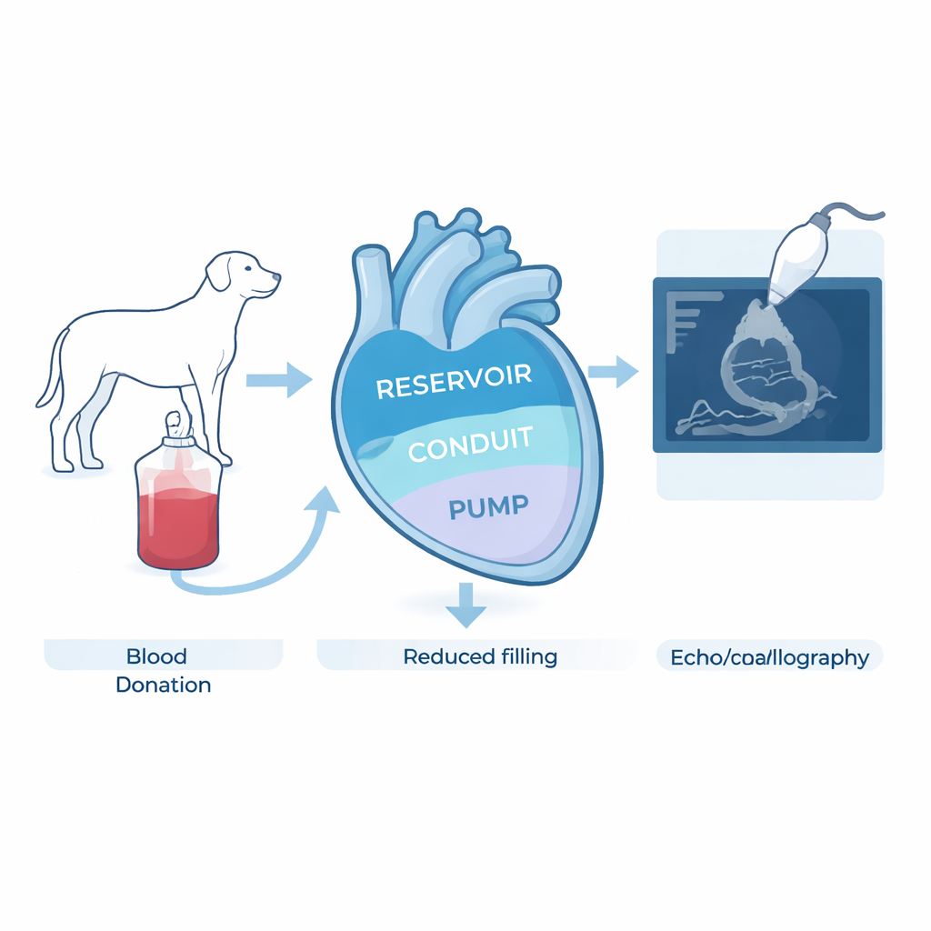
When a dog donates blood, we usually think about the life it might save, not how that donation briefly changes the dog’s own heart. This study looks at what happens inside the heart’s left atrium—the chamber that helps fill the main pumping chamber—right before and after a routine blood donation in healthy dogs. Understanding these subtle changes can make heart ultrasound tests more accurate in both veterinary clinics and, by extension, human medicine.
The heart’s quiet helper chamber
The left atrium is a small but vital part of the heart. It acts in three ways over each heartbeat: first as a storage tank while the main ventricle squeezes, then as a passive channel that lets blood flow forward, and finally as an active booster pump that gives blood an extra push. Doctors and veterinarians can measure how well these phases work using ultrasound scans and special software that tracks how the atrial wall stretches and shortens—a measurement called “strain.” But these numbers are influenced by how much blood returns to the heart (the “preload”), and it has been unclear which measurements truly reflect heart health and which simply mirror momentary changes in blood volume.
Using blood donors as a natural experiment
The researchers worked with 26 healthy, client-owned dogs enrolled in a veterinary blood donor program. All dogs were medium to large breeds, between one and eight years old, and carefully screened to ensure they were free of heart and other systemic diseases. Each dog had a detailed ultrasound exam just before giving blood and immediately afterward, with no sedation or invasive procedures. On average, dogs donated about 11 mL of blood per kilogram of body weight, a volume well within accepted safety limits. This setup turned a real-world clinical act—blood donation—into a controlled, ethical way to study what happens to the heart when circulating volume is suddenly reduced.
What changed in the atrium after blood loss
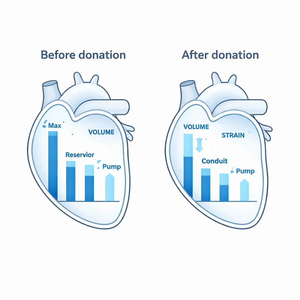
After donation, several key measures of left atrial function dropped. The largest size the atrium reached during the heartbeat (its maximum volume) decreased, as did the volume of blood it ejected and its ejection fraction, a percentage that describes how effectively it empties. Strain readings showed that the atrium’s “storage” role (reservoir strain) and its active squeeze (contractile strain) also went down. Together, these shifts confirmed that many commonly used ultrasound measures of atrial performance are strongly tied to how full the circulation is at the moment of the exam.
The measures that stayed steady
Interestingly, not everything changed. The smallest size of the atrium at the end of filling (minimum volume), the volume just before it actively contracts, and the measure linked to its passive “channel” role (conduit strain) remained essentially stable, even in dogs that donated at least 10 mL/kg. These more stable readings changed little despite a noticeable drop in overall blood volume. That suggests they may be less sensitive to short-term shifts in circulation and could serve as more reliable markers when doctors or veterinarians are trying to judge underlying heart function in patients whose blood volume is changing, such as those who are dehydrated or bleeding.
What this means for heart tests and blood donation
This work shows that most left atrial measurements taken during an ultrasound reflect not only how healthy the heart is, but also how much blood is currently returning to it. For clinicians, this means that volume status must be considered when interpreting certain indices—especially maximum atrial size, how much the atrium empties, and how strongly its wall stretches and contracts. At the same time, the findings highlight a practical, humane way to study heart responses in awake animals: standard canine blood donation. Because it is safe, reproducible, and closely mirrors real clinical situations, this model may help refine noninvasive heart testing in both dogs and humans, ultimately improving how we diagnose and monitor heart disease.
Citation: Cepinho, R.B., de Souza, A.A.L., Reyes, C.J.L. et al. Left atrial strain response to acute preload reduction in healthy dogs using a translational blood donation model. Sci Rep 16, 5479 (2026). https://doi.org/10.1038/s41598-026-35086-4
Keywords: canine cardiology, echocardiography, blood donation, left atrial function, preload