Clear Sky Science · en
Differences of anterior segment features in fellow eyes of primary angle closure glaucoma and healthy eyes
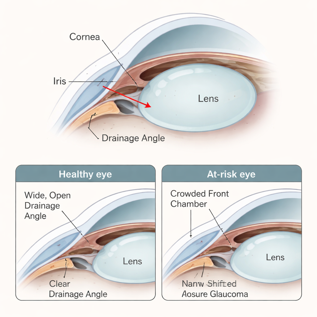
Why the Front of the Eye Matters for Vision
Glaucoma is one of the world’s leading causes of blindness, yet many people at risk feel perfectly fine until serious damage has already occurred. This study looks closely at the front part of the eye in people who are likely to develop a specific form called primary angle-closure glaucoma. By comparing “quiet” eyes in patients who have had problems in only one eye with the eyes of healthy volunteers, the researchers aimed to spot early warning shapes and positions inside the eye that might predict who is headed for a sudden attack versus slow, silent damage.
The Two Ways a Drain Can Clog
Fluid inside the eye normally drains away through a tiny gap where the clear window (cornea) meets the colored part (iris). In primary angle-closure glaucoma, this drainage angle becomes too narrow or even closes, and pressure builds up. This can happen in two main ways. In an acute attack, pressure rises suddenly, causing pain and blurred vision. In the chronic form, the angle slowly narrows over time, quietly harming the optic nerve. The study examined the “fellow eyes” of 72 patients who had glaucoma in only one eye—42 with a history of acute attacks and 30 with chronic disease—and compared them with 22 healthy people. These fellow eyes had not yet been damaged, making them ideal for spotting early structural differences.
Measuring the Eye’s Interior Shape
The team used a high‑resolution ultrasound technique to capture cross‑sectional images of the front of each eye. From these scans, they measured how deep and wide the front chamber of the eye was, how much space it contained, how far forward the natural lens bulged, how curved the iris was, and how thick the iris was near its outer edge. They also calculated how open the drainage angle was, using several precise distance, area, and angle measures. In addition, they introduced a new index describing what fraction of the front eye space was occupied by the lens, hoping this would better capture how “crowded” the front of the eye is.
A Crowded Front Chamber in At-Risk Eyes
Compared with healthy volunteers, both groups of at‑risk eyes—the ones paired with acute attacks and those paired with chronic disease—shared a common theme: the front chamber was shallower and smaller, and the drainage angle was much narrower. At the same time, the lens sat farther forward and occupied more of the limited space. These changes create a crowded environment at the eye’s drainage area, making it easier for the iris to block fluid outflow. However, there were also important differences between the two glaucoma subtypes. Eyes paired with acute attacks tended to have a more forward‑bulging lens and a more bowed or “domed” iris, while eyes paired with chronic disease showed thicker iris tissue at the periphery.
Different Paths to the Same Problem
When the researchers ran statistical tests to see which measurements best separated eyes linked to acute attacks from those linked to chronic damage, one feature stood out: how far the lens bulged forward (called lens vault). A higher lens vault was strongly associated with eyes at risk of an acute pressure spike. In contrast, thickening of the outer iris seemed particularly important in eyes prone to the chronic form, where the drainage angle is gradually crowded and covered. Measures that describe how open the angle is mainly reflected how blocked it already is, rather than acting as early risk factors on their own.

What This Means for Protecting Sight
To a lay reader, the main message is that not all “narrow-angle” eyes are the same. Some eyes are primed for sudden, painful attacks because the lens is pushed too far forward and the iris bulges like a sail, while others quietly lose vision over years because the outer iris is thick and slowly pinches off the drain. By identifying lens position and iris shape in eyes that still see normally, doctors may be better able to decide who needs preventive treatment—such as creating a tiny laser opening in the iris or considering early lens surgery—and who can be monitored more conservatively. Although larger studies are still needed, focusing on how much the lens crowds the front of the eye could become a key step in preventing blindness from angle‑closure glaucoma.
Citation: Guo, L., Wu, Y., Wang, N. et al. Differences of anterior segment features in fellow eyes of primary angle closure glaucoma and healthy eyes. Sci Rep 16, 5135 (2026). https://doi.org/10.1038/s41598-026-35075-7
Keywords: glaucoma, angle-closure, lens position, iris anatomy, eye pressure