Clear Sky Science · en
Computer-aided diagnosis of neonatal acute bilirubin encephalopathy with multi-modal MRI images and convolutional neural networks
Why this matters for newborns and their families
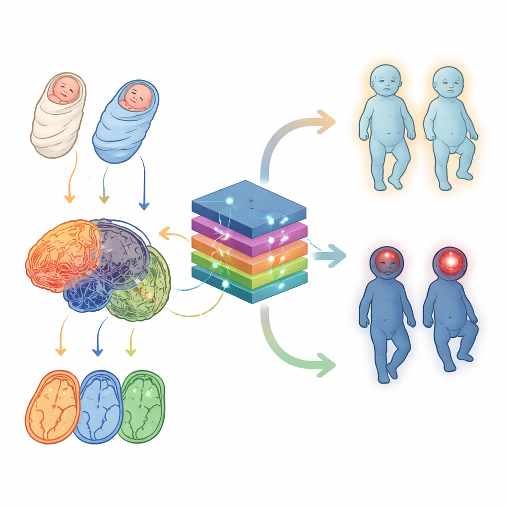
Jaundice is common in newborn babies and is usually harmless, but in some infants a buildup of a yellow pigment called bilirubin can quietly injure the brain. Doctors struggle to tell, from scans and blood tests alone, which jaundiced babies are in real danger. This study explores whether smart computer tools can read brain scans more precisely than the human eye, helping doctors protect babies from permanent disability while avoiding unnecessary alarm and treatment.
The hidden danger behind newborn jaundice
Most babies develop some degree of jaundice as their immature livers learn to clear bilirubin from the blood. When bilirubin levels become very high, the pigment can seep into sensitive deep structures of the brain and trigger a condition called acute bilirubin encephalopathy, which can lead to long-term movement, hearing, and learning problems if not treated in time. The difficulty is that early brain damage can be subtle and reversible, and current bedside scores and lab tests are imperfect. Magnetic resonance imaging (MRI) offers a noninvasive window into the newborn brain, but even specialists often find that affected and unaffected babies look surprisingly similar on standard images.
Looking at the brain with more than one pair of glasses
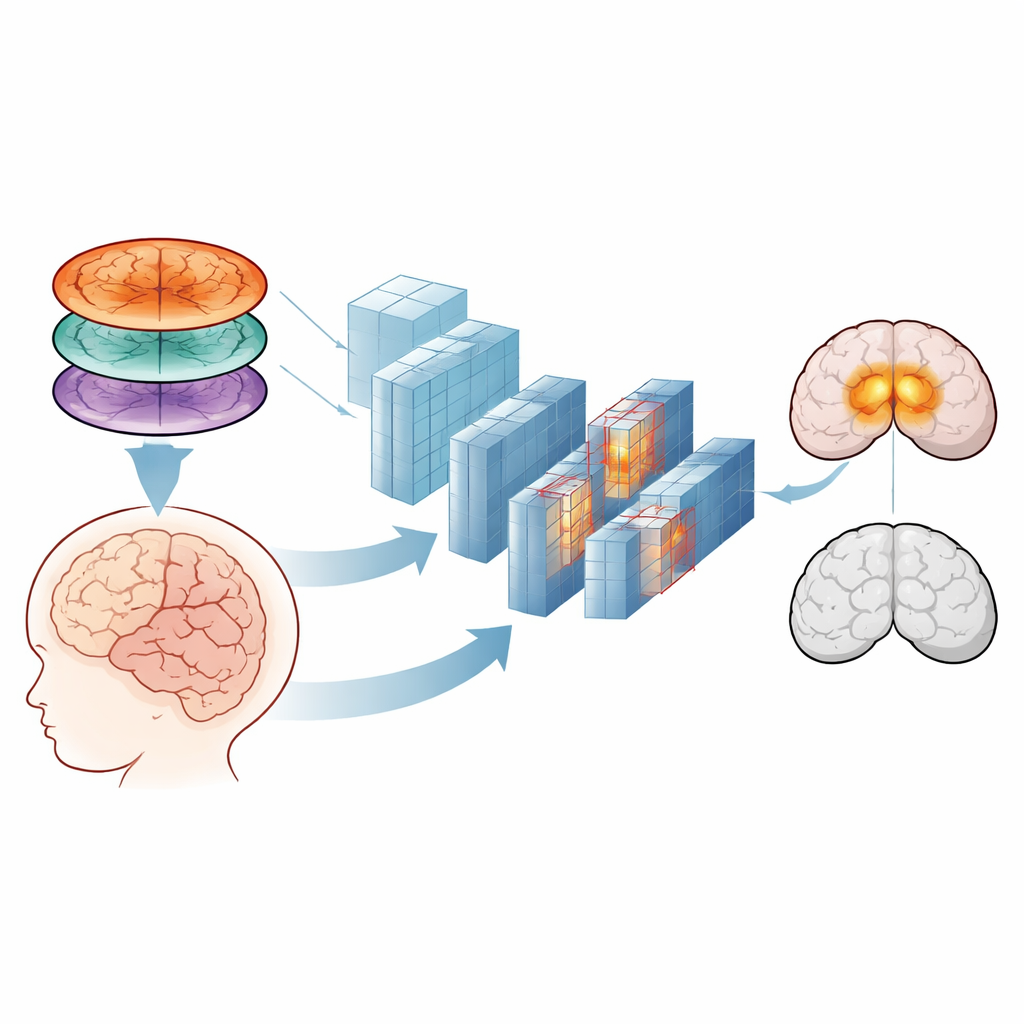
Traditional MRI for these babies focuses mainly on one type of image, known as T1-weighted imaging, where the globus pallidus—a small, deep region involved in movement—can appear unusually bright when injured by bilirubin. Earlier work showed that reading only these images, even with simple computer measurements or earlier deep learning models, left plenty of room for error. The authors reasoned that other MRI “flavors,” such as T2-weighted images and diffusion-based maps that trace how water moves through brain tissue, might contain extra clues. Instead of asking doctors to manually measure specific regions, they set out to feed the full richness of these three image types into modern image-recognition algorithms.
Teaching computers to spot early brain injury
The research team collected brain scans from 150 newborns with high bilirubin levels: half had signs of acute brain involvement and half did not. For each baby, three MRI series were obtained—T1, T2, and a diffusion-based map called an apparent diffusion coefficient (ADC). First, the images were carefully aligned and cropped so only the brain remained, then resized and brightness-normalized so they could be handled by standard image-analysis software. The scientists tried two broad strategies. One relied on a classic machine-learning method called a support vector machine, using simple hand-calculated brightness ratios between a vulnerable deep structure (the globus pallidus) and nearby white matter. The other used powerful deep learning models, InceptionV3 and EfficientNetB0, which learn their own visual features directly from the raw images.
Mixing scan types gives the clearest picture
When the team compared methods, the simpler, hand-measured approach did improve somewhat when multiple scan types were combined, but its best performance still fell short of what would be comfortable for clinical decision-making. In contrast, the deep learning models improved dramatically as more MRI types were fused. By stacking T1, T2, and ADC images together like three color channels in a photograph, the best-performing network (InceptionV3) correctly distinguished affected from unaffected babies in more than 96% of cases, with an almost perfect measure of overall discriminative power. Visual maps of which regions the network relied on showed that it focused on the same deep brain areas—globus pallidus, subthalamic nuclei, and hippocampus—that human experts consider most vulnerable to bilirubin injury, suggesting that the computer was learning clinically meaningful patterns rather than random quirks.
From research tool to bedside helper
The study concludes that a carefully trained computer-aided diagnosis system, fed with several complementary types of MRI rather than a single scan, can flag early bilirubin-related brain injury in newborns with impressive accuracy. While the work was done at a single hospital and still faces hurdles such as overfitting and the need to test on larger, more diverse groups of babies, it points toward a future in which advanced image-reading algorithms act as a second set of eyes for pediatricians and radiologists. Used wisely, such tools could help identify which jaundiced infants most urgently need treatment—and just as importantly, reassure families when a baby’s brain is likely to be safe.
Citation: Wu, M., Liu, Q. Computer-aided diagnosis of neonatal acute bilirubin encephalopathy with multi-modal MRI images and convolutional neural networks. Sci Rep 16, 9611 (2026). https://doi.org/10.1038/s41598-025-33337-4
Keywords: neonatal jaundice, bilirubin encephalopathy, brain MRI, deep learning, computer-aided diagnosis