Clear Sky Science · en
Early detection of retinal changes in children with diabetes without retinopathy using optical coherence tomography and optical coherence tomography angiography
Why children’s eye health matters in diabetes
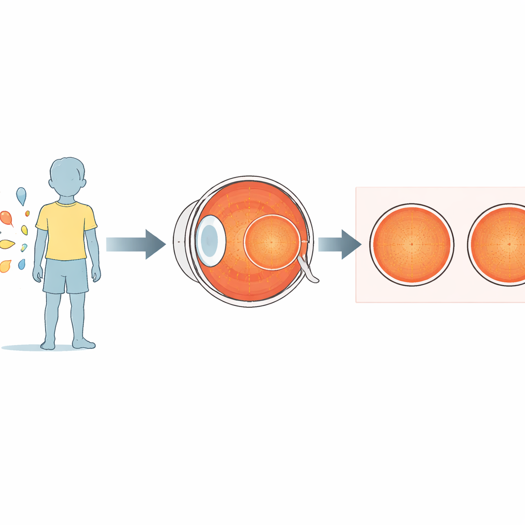
For many families, a diagnosis of diabetes in a child raises urgent questions about long-term health, including the risk of vision loss. The light-sensitive tissue at the back of the eye, the retina, can be damaged slowly and silently for years before standard eye exams detect any problem. This study explores whether modern, non-invasive imaging tools can reveal the earliest hidden changes in the eyes of children and teenagers with diabetes—even when their retinas still look completely normal in routine checks.
Looking for hidden warning signs in young eyes
Diabetic retinopathy, the eye disease caused by long-term high blood sugar, is a leading cause of blindness in adults. Historically, it has been considered uncommon and rarely sight-threatening in children, which is why many screening guidelines only recommend detailed eye exams several years after diagnosis. However, rising rates of both type 1 and type 2 diabetes in young people have prompted researchers to ask whether subtle retinal damage might begin earlier than expected. Detecting these changes before symptoms appear could allow doctors to better time eye screening and intervene earlier to protect vision.
High-tech scans beyond the standard eye exam
The research team in Bangkok studied 300 participants aged 8 to 18 years: 148 with diabetes and 150 healthy children matched for age and sex. None of the children with diabetes showed visible signs of diabetic retinopathy on ultra-widefield photographs of the back of the eye. In addition to standard eye checks, the researchers used two advanced, painless imaging techniques. Optical coherence tomography (OCT) works a bit like an optical ultrasound, building highly detailed cross-sections of the retina and the underlying layer that supplies it with blood. Optical coherence tomography angiography (OCTA) goes a step further by mapping tiny blood vessels and the fine network of capillaries that nourish the central part of vision.
Subtle thinning and reduced blood supply around the center of vision
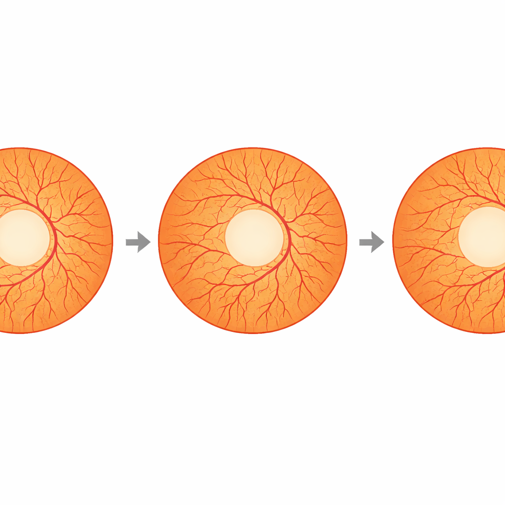
When the team compared the scans from children with diabetes to those from healthy controls, most overall measurements looked similar. Central retinal thickness and the thickness of the deeper vascular layer were not significantly different. But a more focused look at the band of tissue just around the very center of the retina—the parafoveal zone—revealed that this region was slightly thinner in the diabetes group in several directions (temporal, superior, and nasal). On the blood-flow maps from OCTA, a particularly sensitive measure stood out: the density of tiny vessels in a narrow ring around the small vessel-free spot at the center of the retina. This ring, called FD-300 in the study, was measurably lower in children with diabetes than in healthy peers, even though their eyes still appeared normal in regular photographs.
Changes that grow with years of diabetes
The researchers then asked what might be driving these early changes. They grouped the children with diabetes by how long they had been living with the disease and by their long-term blood sugar control. Children who had diabetes for more than five years showed the greatest drop in vessel density around the foveal center and also some reduction in blood vessel density in the surrounding zone. Statistical analysis showed that the longer a child had diabetes, the lower this ring-shaped vessel density tended to be, and the thinner a specific part of the parafoveal retina became. By contrast, the study found only limited and less consistent links between these eye measurements and average blood sugar over time, suggesting that duration of disease may be a particularly important driver of early retinal change in this young population.
What this means for families and doctors
Overall, the study suggests that the eyes of children and teenagers with diabetes can show measurable structural and blood-flow changes long before traditional signs of diabetic retinopathy appear. A carefully measured reduction in vessel density around the center of vision—and slight thinning of the nearby retinal tissue—may act as early, non-invasive warning markers of future disease. For families, this does not mean that vision loss is inevitable, but it underscores the value of regular, appropriately timed eye imaging in addition to routine exams. For clinicians and researchers, these findings point to FD-300 and related measures as promising tools to detect silent retinal stress early, refine screening guidelines for young patients, and ultimately help preserve sight over a lifetime with diabetes.
Citation: Montrisuksirikun, C., Santiprabhob, J., Lertbannaphong, O. et al. Early detection of retinal changes in children with diabetes without retinopathy using optical coherence tomography and optical coherence tomography angiography. Sci Rep 16, 8457 (2026). https://doi.org/10.1038/s41598-025-31609-7
Keywords: pediatric diabetes, diabetic retinopathy, retinal imaging, OCT angiography, early eye changes