Clear Sky Science · en
A two-stage deep learning framework for kidney disease detection using modified specular-free imaging and EfficientNetB2
Why clearer kidney scans matter
Kidney diseases such as tumors, cysts, and stones affect hundreds of millions of people worldwide, and catching them early can change the course of a person’s life. Doctors often rely on CT scans to spot these problems, but the images can be hard to read: glare, shadows, and low contrast can hide small but important details. This study presents a new computer-based system that first cleans up kidney images and then uses artificial intelligence to tell healthy kidneys from diseased ones with very high accuracy.
Making tricky scans easier to read
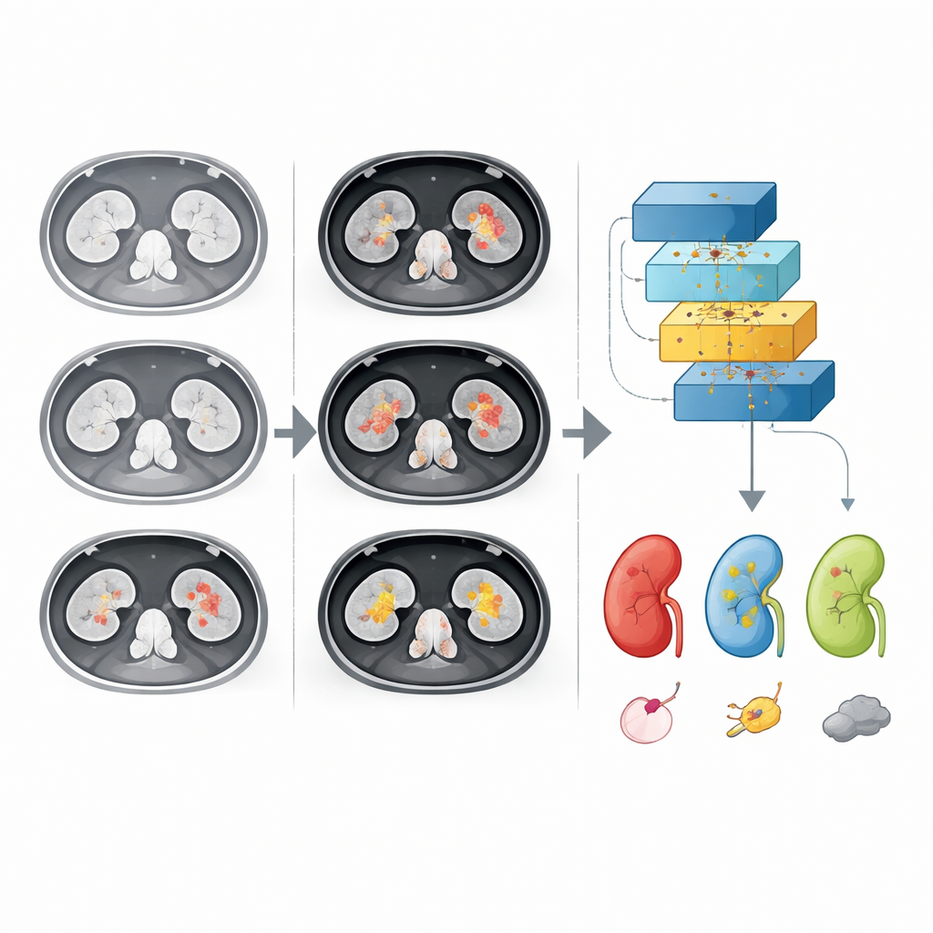
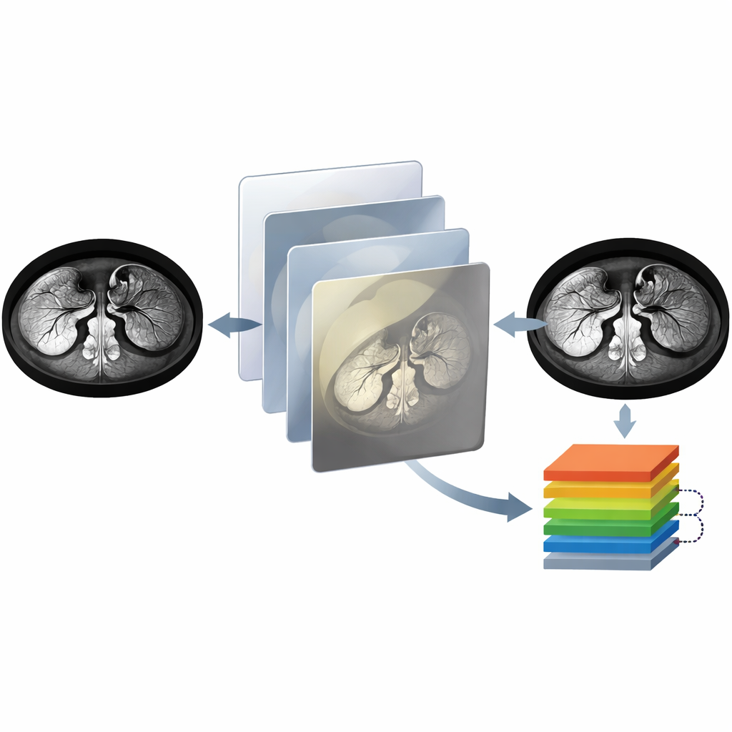
Medical CT images are rarely perfect. Shiny reflections from bone or surgical clips, uneven lighting, and washed-out or overly dark regions can all blur the line between normal tissue and a hidden tumor or stone. The authors tackle this by designing a special enhancement method called Modified Specular-Free imaging. Instead of simply boosting the overall contrast, their method looks at the color and brightness of each pixel to remove glare-like highlights while preserving the true structure and shading of soft tissues. It then decides whether an image is mostly dark or mostly bright and adjusts it differently in each case, so both shadowed and overexposed areas become easier to interpret.
Turning blurry data into sharp detail
After the initial clean-up, the system refines the image further using a technique that estimates how light falls across the scene. This step brightens dim regions and tones down overly bright ones, creating a more balanced picture of the kidneys. A high‑dynamic‑range style adjustment then stretches the range of visible detail so that subtle differences inside the kidney stand out instead of blending into the background. Together, these steps produce CT images in which the borders of cysts, stones, and tumors become clearer, and small abnormalities that might have been missed are easier to see. The team also checks that these enhancements truly add useful information by measuring how much structure and natural appearance the images retain.
Letting a smart network read the scans
Once the images are enhanced, they are passed to a modern deep learning model known as EfficientNet‑B2. This model is built from many layers of simple pattern detectors that gradually learn to recognize edges, textures, and higher‑level shapes in the kidneys. Trained on more than twelve thousand labeled CT images—spanning normal kidneys, cysts, tumors, and stones—the network learns which visual patterns go with each diagnosis. The authors compare this model with several well‑known neural networks and transformer‑based systems and find that EfficientNet‑B2, combined with their enhancement pipeline, delivers a powerful mix of accuracy and speed that is practical for hospital use.
How well the system performs
On unseen test images, the two‑stage system correctly identifies the type of kidney condition in the vast majority of cases, reaching an accuracy of about 98%. It not only outperforms classic deep learning models like VGG and ResNet in this task, but also rivals or surpasses newer, more computationally heavy designs. The researchers show that their enhancement steps alone boost performance by several percentage points, confirming that cleaning the images is just as important as the choice of network. They also analyze where the system still makes mistakes—such as confusing a normal kidney with a tumor when textures look similar—and suggest that adding more diverse training examples and fine‑tuning the enhancement could reduce these errors further.
What this means for patients and clinics
In plain terms, this work shows that pairing smarter image cleaning with an efficient deep learning model can help computers spot kidney problems on CT scans both accurately and quickly. While it does not replace radiologists, such a tool could act as an always‑alert assistant: flagging suspicious cases, supporting diagnoses in busy clinics, and potentially making advanced kidney care more accessible in settings with limited specialist expertise. With broader testing on more diverse patient groups and better ways to explain its decisions to doctors, this type of system could become a routine part of how kidney diseases are detected and monitored.
Citation: El-Hag, N.A., El-Shafai, W., El-Hameed, H.A.A. et al. A two-stage deep learning framework for kidney disease detection using modified specular-free imaging and EfficientNetB2. Sci Rep 16, 8358 (2026). https://doi.org/10.1038/s41598-025-04606-z
Keywords: kidney CT, deep learning, medical imaging, kidney tumors and stones, image enhancement