Clear Sky Science · en
Light sheet microscopy imaging dataset of CAR-T-cell-mediated cytotoxicity
Watching Cancer-Fighting Cells in Action
Cancer therapies that enlist our own immune system, such as CAR-T cells, are transforming medicine, but researchers still struggle to watch exactly how these living drugs battle tumor cells in real time. This study introduces a powerful new imaging dataset and microscope system that let scientists follow hundreds of individual cancer-fighting encounters in 3D, for hours at a stretch, without harming the cells with light. The freely available data are designed to accelerate discoveries about why some immune cells wipe out tumors while others stall.
A New Window on Living Cancer Killers
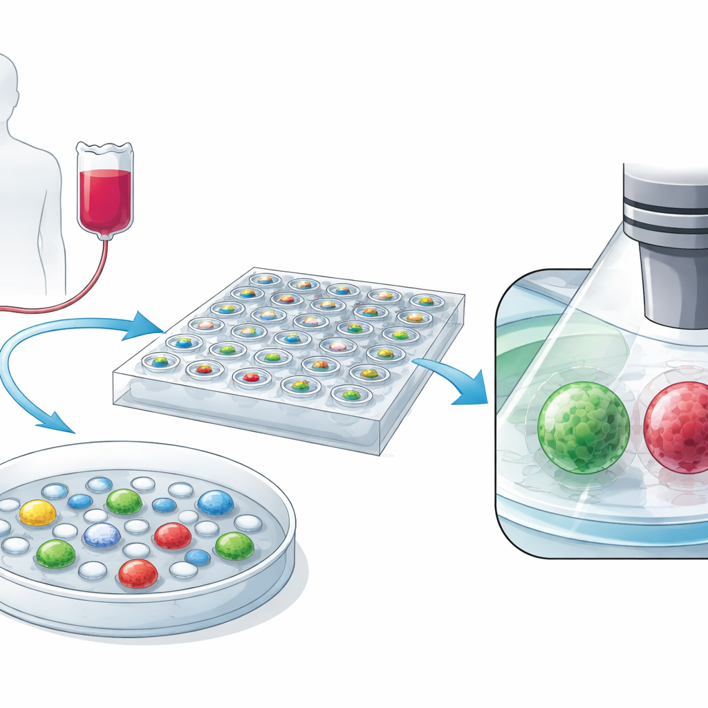
CAR-T cells are a patient’s own T cells that have been reprogrammed to recognize cancer. How well they work depends on their moment-to-moment behavior: how they move, grab onto a target, and deliver the fatal blow. Traditional microscopes can zoom in on these events but often damage delicate cells with intense light and cannot keep up with fast changes or long experiments. The authors set out to close this gap by creating both a new microscope setup and a large, sharable collection of movies that follow CAR-T cells as they interact with leukemia cells over several hours.
Trapping Tiny Duels in Thousands of Mini Wells
To reliably watch many one-on-one battles, the team first had to keep floating cells from drifting away under the microscope. They built a transparent microchip that contains 2,025 tiny cylindrical wells, each about the width of a human hair. CAR-T cells and leukemia cells are mixed together and gently allowed to settle into these wells, where a simple mathematical model predicts how often a single CAR-T lines up with a single target. The chip material is carefully matched to the refractive index of water so that light passes cleanly, preserving image sharpness across all wells.
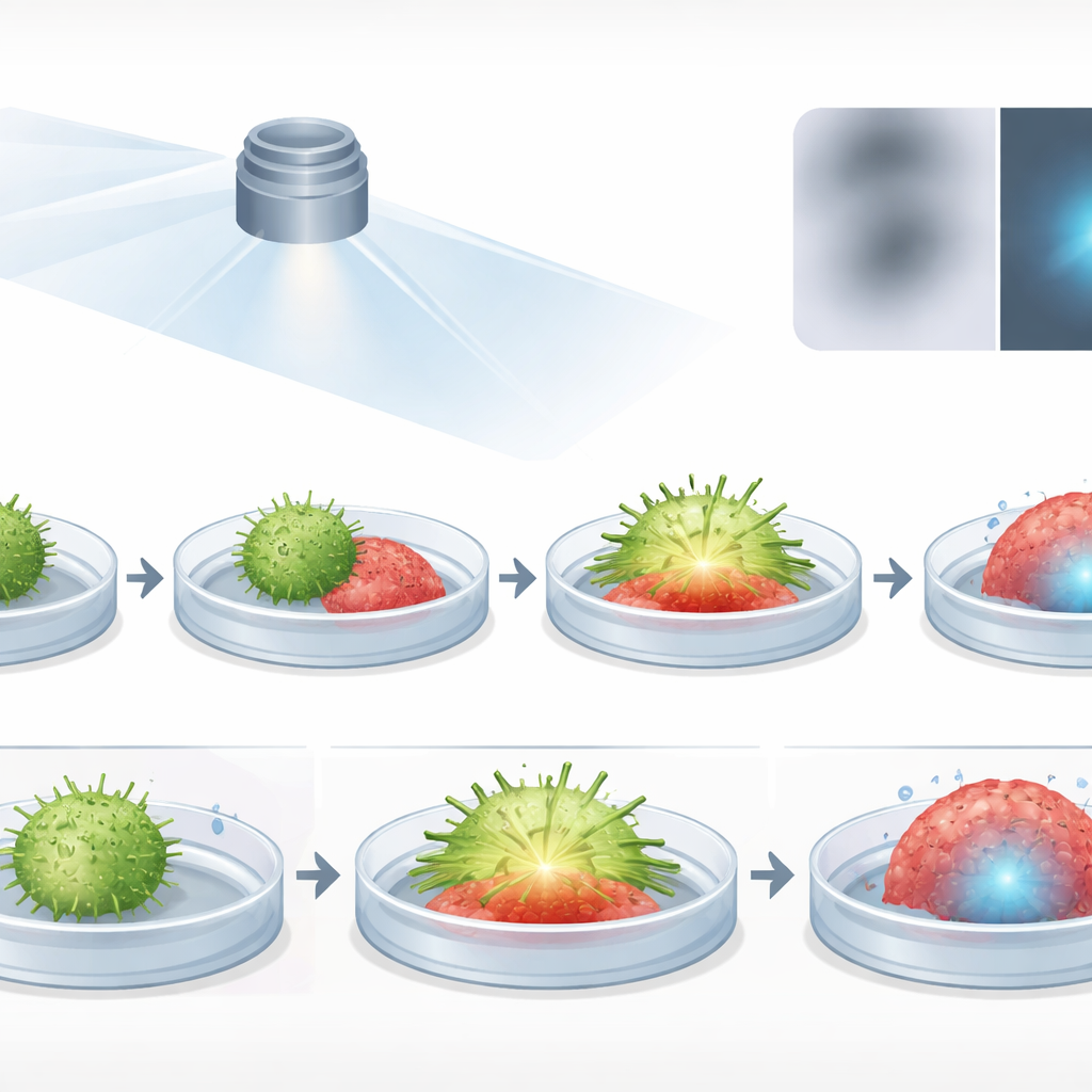
Fast, Gentle 3D Movies of Cell Combat
The heart of the system is a customized light-sheet microscope called high-throughput Bessel oblique plane microscopy. Instead of bathing the whole sample in light, a thin sheet sweeps through the well at an angle, exciting only a narrow slice at a time. Paired with an optical trick that reshapes the images back into an upright 3D volume, this design captures the full shape and internal details of both CAR-T and tumor cells with a resolution of about 320 nanometers. Smart software first scans the chip at low magnification to spot promising cell pairs, then automatically revisits those wells with high magnification to record rapid, repeated 3D stacks while limiting light exposure.
Rich, Color-Coded Data for the Community
The resulting dataset contains more than 400 time-lapse image sets from healthy donors, plus additional sets where CAR-T cells were treated with a drug known to turn down their killing ability. Different fluorescent colors mark the CAR-T cell skeleton, the tumor cell membrane, the internal scaffolding of CAR-T cells, and the nuclei of cells that have died. The authors provide not only the raw image files but also reconstructed 3D volumes and machine-generated outlines that separate immune cells, tumor cells, and their nuclei. A graphical interface helps users reprocess volumes, adjust noise, and pull out specific time points or channels for further analysis.
Proving It Works and Why It Matters
To test the system, the researchers compared it with a standard confocal microscope and found that their approach can record roughly 50 times more 3D volumes before the signal fades to the same level, confirming far lower light damage. They also showed that the images faithfully capture known biology: CAR-T cells exposed to an inhibitory drug form smaller contact zones with tumors, move their internal scaffolding more slowly, and kill fewer target cells, just as expected. Together, the microscope design and open dataset give scientists a powerful new way to watch living cancer therapies at work and to uncover what makes some cells potent tumor killers—and how future treatments might make more of them behave that way.
Citation: Wang, J., Jin, J., Fang, Y. et al. Light sheet microscopy imaging dataset of CAR-T-cell-mediated cytotoxicity. Sci Data 13, 439 (2026). https://doi.org/10.1038/s41597-026-06829-9
Keywords: CAR-T cells, light-sheet microscopy, cancer immunotherapy, live-cell imaging, single-cell dynamics