Clear Sky Science · en
BRISC: Annotated Dataset for Brain Tumor Segmentation and Classification
Why Brain Scan Data Matters for Everyone
Brain tumors are among the most frightening diagnoses a person can receive, and doctors increasingly rely on computer programs to help spot and outline these dangerous growths in MRI scans. But like students learning from a textbook with missing pages, many of today’s artificial intelligence (AI) systems are held back by incomplete or inconsistent data. This article introduces BRISC, a new carefully assembled collection of brain MRI images created to give medical AI the high‑quality examples it needs to better detect and map brain tumors—work that could ultimately support faster, more reliable diagnoses. 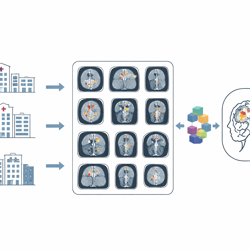
A New Library of Brain Images
The BRISC dataset gathers 6,000 MRI brain images focused on a specific kind of scan—contrast‑enhanced T1‑weighted images—which are particularly good at making tumor edges stand out. Each image falls into one of four groups: three common tumor types (glioma, meningioma, and pituitary tumors) plus a non‑tumorous group that includes healthy brains and other non‑cancerous conditions. The pictures come from several earlier public collections, but BRISC adds what those older sets largely lacked: precise outlines of the tumor regions and consistent labels, created and checked by medical experts.
Balancing Views and Types of Tumors
One major problem with many existing collections is imbalance: some tumor types or scan angles dominate, nudging AI models to perform well only on the most common patterns they see. BRISC tackles this by designing a more even spread of both diagnoses and viewing directions. Images are provided across three standard MRI views—axial (top‑down), coronal (front‑to‑back), and sagittal (side‑to‑side)—with similar numbers in each. The four diagnosis categories are also kept relatively balanced in the training and test splits. This careful design helps future algorithms learn to recognize tumors from multiple angles and in a broader range of situations, better reflecting what doctors actually see in clinics.
Careful Cleaning and Expert Outlining
Turning raw scans into a trustworthy research resource required substantial cleanup. The team began with more than 7,000 images taken from a popular online brain‑tumor collection and removed poor‑quality or corrupted scans, near‑duplicate images, and sequences too short for reliable interpretation. Only contrast‑enhanced T1 scans were kept to maintain consistency. Physicians and a radiologist then reviewed the images, correcting wrong labels and removing questionable cases. Using a specialized labeling tool, they drew detailed masks around tumor regions, repeatedly refining their work until they reached strong agreement; on a test subset, the match between initial and expert‑approved outlines was very high. 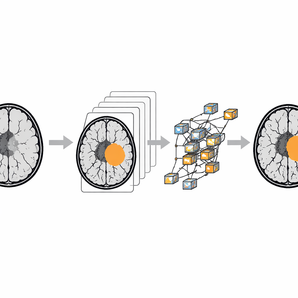
What the Data Enables for AI Models
To show how BRISC can be used, the authors trained a range of popular AI models on two tasks. The first task asks a model to classify each image into one of the four diagnosis categories. Modern image‑recognition systems, especially the EfficientNet family, achieved very high accuracy—correctly labeling the vast majority of scans and performing particularly well at distinguishing images with no tumor. The second task asks models to color in the tumor area, pixel by pixel, on the MRI slice. Here, more advanced segmentation networks, including transformer‑based architectures that excel at modeling context, delivered the best scores, accurately outlining tumors across the three main tumor types.
How This Work Moves the Field Forward
In plain terms, BRISC is a well‑organized, public “training ground” for computers learning to read brain MRIs. It offers thousands of carefully cleaned scans, realistic variety across tumor types and viewing angles, and expert‑drawn tumor outlines that teach algorithms exactly where disease is present. While the dataset is intended for research—not as a stand‑alone diagnostic tool for patients—it provides a solid foundation for building and comparing new AI systems. As researchers refine models using BRISC and similar resources, doctors may one day have more dependable digital assistants at their side, helping them detect brain tumors earlier and plan treatments with greater confidence.
Citation: Fateh, A., Rezvani, Y., Moayedi, S. et al. BRISC: Annotated Dataset for Brain Tumor Segmentation and Classification. Sci Data 13, 361 (2026). https://doi.org/10.1038/s41597-026-06753-y
Keywords: brain tumor MRI, medical imaging AI, tumor segmentation, dataset curation, radiology deep learning