Clear Sky Science · en
An EEG dataset with carbon wire loops in cognitive tasks and resting state inside and outside MR scanners
Why Cleaner Brain Scans Matter
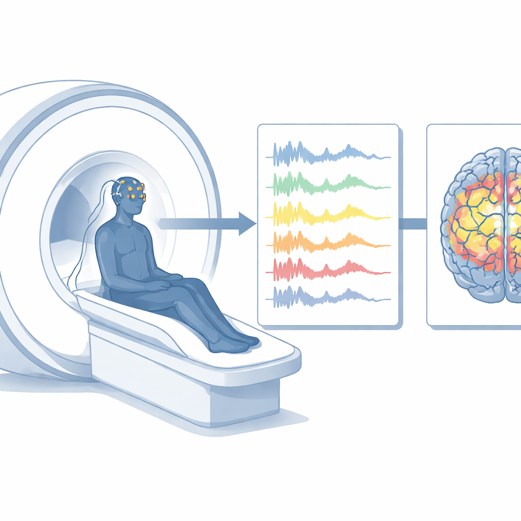
Brain scanners and brainwave caps let scientists watch our minds at work, but combining these tools is surprisingly messy. Magnetic resonance imaging (MRI) reveals where activity happens deep inside the brain, while electroencephalography (EEG) tracks split‑second electrical signals at the scalp. When both are used at the same time, powerful magnets and tiny body movements flood the EEG with noise, obscuring the very signals researchers care about. This study introduces a carefully designed dataset that tackles that problem head‑on, offering a cleaner, more realistic view of brain activity during rest and everyday thinking tasks.
Two Windows on the Working Brain
The researchers recorded brain activity from 39 healthy adults using EEG and functional MRI while people rested with eyes open and performed two simple mental tasks. In a “visual oddball” game, volunteers watched frequent circles and rare stars and silently counted how often the rare shape appeared. In an “N‑back” game, they saw a stream of numbers and pressed a button when a target number appeared immediately (easy version) or matched the one shown two steps earlier (hard version). These tasks are common tools for probing attention and working memory, making the data useful to many labs worldwide.
Inside and Outside the Scanner
Crucially, each person completed these tasks both inside an MRI scanner and in a quiet, shielded room where only EEG was recorded. This pairing allows scientists to ask a fundamental question: how much do the noisy conditions inside the scanner change the signals we observe at the scalp? The team also used two different MRI machines for a subset of participants, creating a “traveling subject” design that helps researchers compare how hardware differences affect the data. All recordings were organized in a standard, machine‑readable format so that other groups can plug the files directly into modern analysis pipelines.
Loops that Listen to Noise
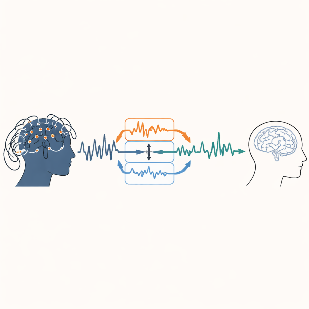
To tame the scanner’s interference, the team relied on an elegant trick: carbon wire loops sewn into the EEG caps. These tiny loops act like dedicated noise microphones, picking up motion‑related disturbances and the subtle pulse‑driven jolts produced when blood moves through the magnetic field. By mathematically subtracting these loop signals from the EEG, the researchers could peel away much of the unwanted noise without damaging the underlying brain activity. They combined this with established cleaning steps, such as filtering, automatic detection of bad channels, and removal of eye‑movement and power‑line artifacts.
Putting Signal Quality to the Test
To check that their cleaning worked, the team looked at the strength of electrical rhythms across frequencies and at time‑locked responses to specific events. After correction, EEG recordings collected inside the scanner resembled those from the quiet room: the strongest scanner‑related ripples were largely gone, while familiar features such as the P300 response—an electrical spike tied to noticing rare or important stimuli—remained visible in both the visual oddball and N‑back tasks. At the same time, the MRI data showed robust and anatomically sensible patterns of activation in brain regions known to support attention and working memory, such as parts of the frontal cortex, parietal cortex, and cerebellum. Differences between the two scanners were mostly in signal strength, not in which regions lit up.
A Sharper Tool for Future Brain Research
In plain terms, this work delivers a well‑documented, publicly accessible dataset that shows it is possible to record brainwaves and brain images together without drowning in noise. By pairing inside‑scanner and outside‑scanner recordings, adding measurements from two MRI systems, and using carbon wire loops to track and cancel unwanted interference, the authors provide a practical blueprint for cleaner multimodal brain studies. Researchers can now use these shared data to test new analysis methods, compare hardware, or explore how attention and memory unfold in the brain, all with greater confidence that the signals they see reflect real neural activity rather than the heartbeat of the machine.
Citation: Tsutsumi, M., Kishi, T., Ogawa, T. et al. An EEG dataset with carbon wire loops in cognitive tasks and resting state inside and outside MR scanners. Sci Data 13, 351 (2026). https://doi.org/10.1038/s41597-026-06734-1
Keywords: simultaneous EEG fMRI, brain imaging dataset, artifact reduction, working memory, attention