Clear Sky Science · en
RVO-ME: A Dual-Task OCT Dataset for Segmentation and Detection of Macular Lesions in Retinal Vein Occlusion
Why this matters for eyesight
When a major vein in the back of the eye becomes blocked, vision can blur or disappear, often without warning. Doctors now rely on a powerful imaging technique, optical coherence tomography (OCT), to see swelling and damage in the retina. This article introduces a carefully built image collection that helps computers learn to read those scans, with the long‑term goal of faster, more accurate diagnosis and treatment planning for people at risk of losing their sight.
A common cause of sudden vision loss
Retinal vein occlusion is one of the leading blood‑vessel diseases of the eye, affecting an estimated 28 million people worldwide. When a retinal vein is blocked, fluid leaks into the central part of the retina, the macula, causing macular edema and blurred vision. Drugs that block a signaling molecule called VEGF have greatly improved treatment, but not all patients respond well. Doctors therefore look for subtle signs in OCT scans that can predict who is likely to benefit most and how vision will change over time. Until now, progress in using artificial intelligence to read these scans has been slowed by a simple problem: there were not enough high‑quality, expertly labeled images focused specifically on this disease.
Building a detailed picture library
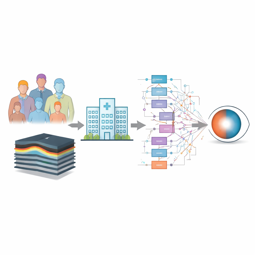
The research team created a new dataset called RVO‑ME, made from 3,012 OCT cross‑section images of the macula collected from 146 eyes of 130 patients at a single hospital in China between 2019 and 2024. Every image went through strict screening to exclude poor quality scans or eyes with other serious retinal conditions. All personal information was removed, and patients gave written consent for their images to be used in research and in a public data resource. The scans capture the retina both before and after treatment, giving a broad view of how the disease and its complications appear in everyday clinical practice.
Marking the tiny clues in each scan
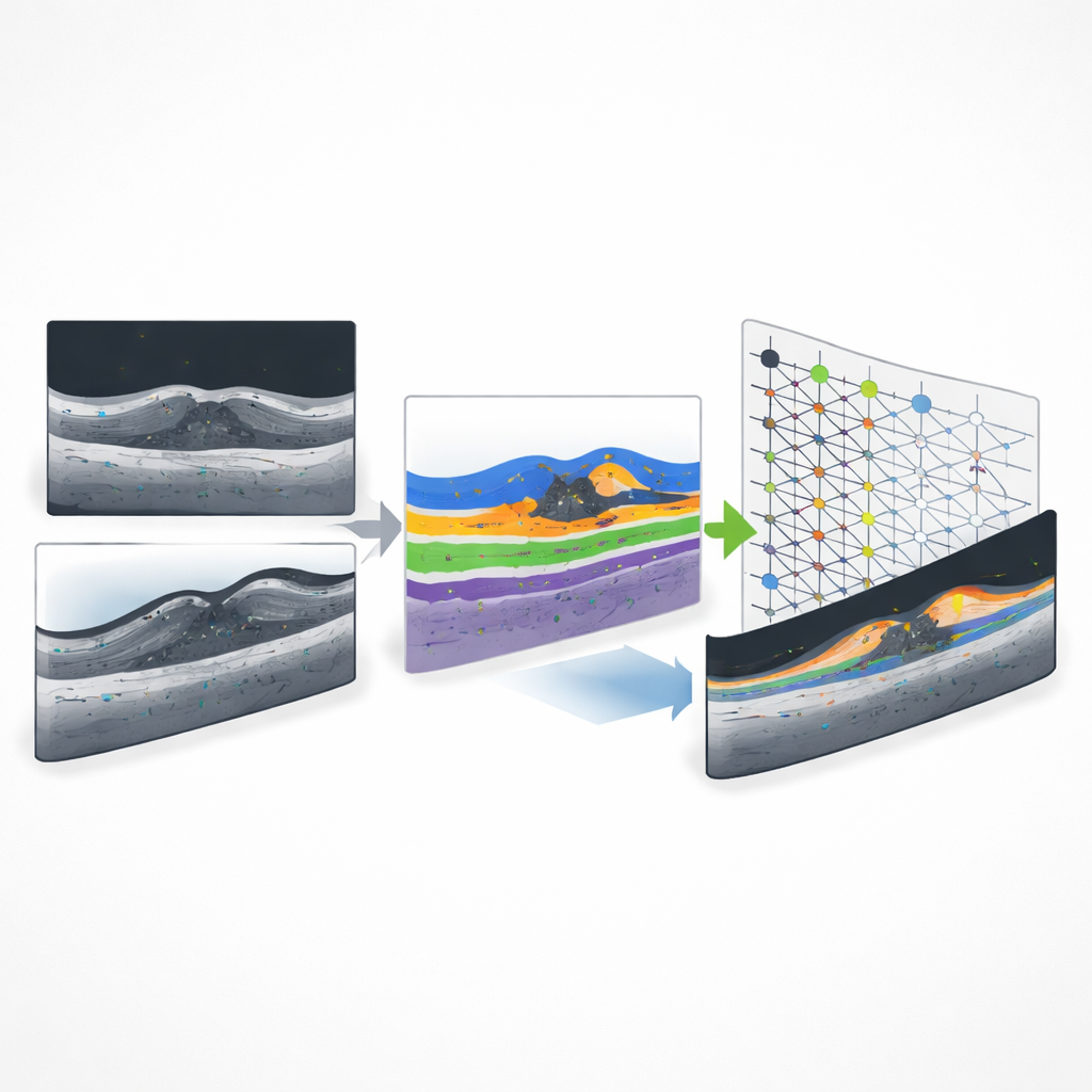
To turn this picture library into a training ground for computers, the authors needed to trace, by hand, the key signs that matter most for vision. Three junior ophthalmologists used specialized software to outline pockets of fluid inside and under the retina, draw thin lines marking two important light‑reflecting bands, and place points on tiny bright specks known as hyperreflective foci. These markings were then checked and corrected by a senior retinal specialist, who graded each set of labels and sent back lower‑quality attempts for revision. Before starting at scale, the team ran a consistency exercise in which trainees labeled the same images on different days, confirming that their markings largely agreed, especially for the larger fluid areas. Extra training was focused on the more delicate, thread‑like bands that are easily blurred in diseased eyes.
From expert markings to smart machines
In the final dataset, each OCT image has a matching “mask” image in which every pixel belongs either to background or to one of four key structures, and each tiny bright point is stored for detection tasks. The authors split the images into separate groups for training and testing so that the same patient never appears in both, preventing computers from simply memorizing individual eyes. They then tested several popular image‑analysis algorithms on this collection. For the larger fluid regions, modern segmentation models achieved solid accuracy, with a U‑Net++ approach performing best overall. For the pinpoint bright spots, a more complex two‑stage detection method (Faster‑RCNN) far outperformed a faster, one‑stage model, reflecting how challenging it is to find minute features scattered across noisy medical images.
How this resource could shape future eye care
This dataset does not by itself change how patients are treated, and it has limitations: all scans come from a single type of OCT machine and from patients of one ethnic background. Yet it fills a crucial gap: until now there was no public macular OCT collection dedicated to vein‑related swelling that captured both fluid pockets and fine retinal structures together with tiny bright specks. By making the images, expert markings, and example computer‑analysis code openly available, the authors provide a common reference point for researchers worldwide. Better algorithms trained on such data could one day help eye doctors quickly measure disease severity, predict which patients will benefit most from injections, and track recovery more precisely, ultimately supporting more personalized and efficient care for people facing vision loss from retinal vein occlusion.
Citation: Xiong, F., Li, G., Gao, W. et al. RVO-ME: A Dual-Task OCT Dataset for Segmentation and Detection of Macular Lesions in Retinal Vein Occlusion. Sci Data 13, 349 (2026). https://doi.org/10.1038/s41597-026-06695-5
Keywords: retinal vein occlusion, macular edema, optical coherence tomography, medical imaging dataset, artificial intelligence in ophthalmology