Clear Sky Science · en
Complex multi-echo fMRI dataset: New strategies in processing of multi-echo data
Why this brain scan dataset matters
Modern brain scanners can record not just where activity happens in the brain, but also how that signal changes in subtle ways over time. Yet many studies still use relatively simple scanning methods, leaving a lot of this rich information untapped. This article introduces a carefully designed, openly shared brain imaging dataset that pushes the limits of what functional MRI (fMRI) can do. It is meant as a testbed for new analysis tricks that could make future brain studies more reliable, more detailed, and less noisy.
Looking at the brain from several angles at once
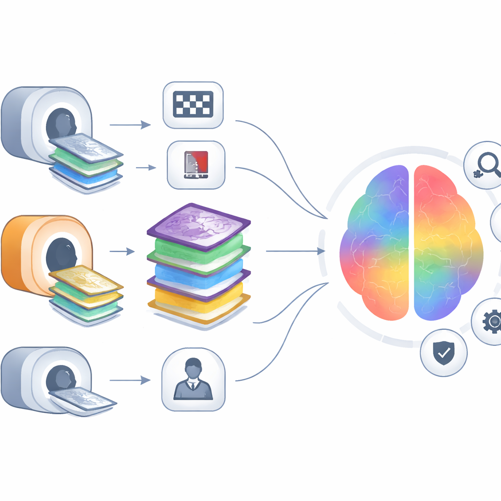
Most fMRI experiments collect one image of the brain every time the scanner “listens” for a signal. In this project, the researchers did something more ambitious: they used a technique called multi-echo fMRI, which captures several images in rapid succession after each scanner pulse. Each of these “echoes” emphasizes different aspects of the signal, including how sensitive it is to blood oxygen changes and how strongly it is affected by distortions and noise. On top of that, the team kept not only the usual signal strength (magnitude) but also the often-ignored phase information, which tracks how the signal’s timing shifts in the magnetic field. That extra phase layer can reveal breathing and heartbeat effects, as well as large veins that might otherwise be mistaken for true brain activity.
A rich mix of tasks, scanners, and signals
The dataset includes 83 healthy adults who lay in a 3‑Tesla MRI scanner and completed six different runs in a single session. They performed three kinds of conditions: a simple visual-and-motor task where they watched a flickering checkerboard and pressed buttons, a more mentally demanding “oddball” task where they responded to rare visual targets among frequent non-targets, and a quiet resting period with eyes closed. Each of these was repeated with two different rhythms of data collection, one slower and one faster, and all of it was recorded on two nearly identical scanners that differed only in a few timing and hardware settings. Alongside the brain images, the team also stored high-quality heart and breathing traces, plus extra structural scans and field maps that help correct for distortions.
Building a playground for better methods
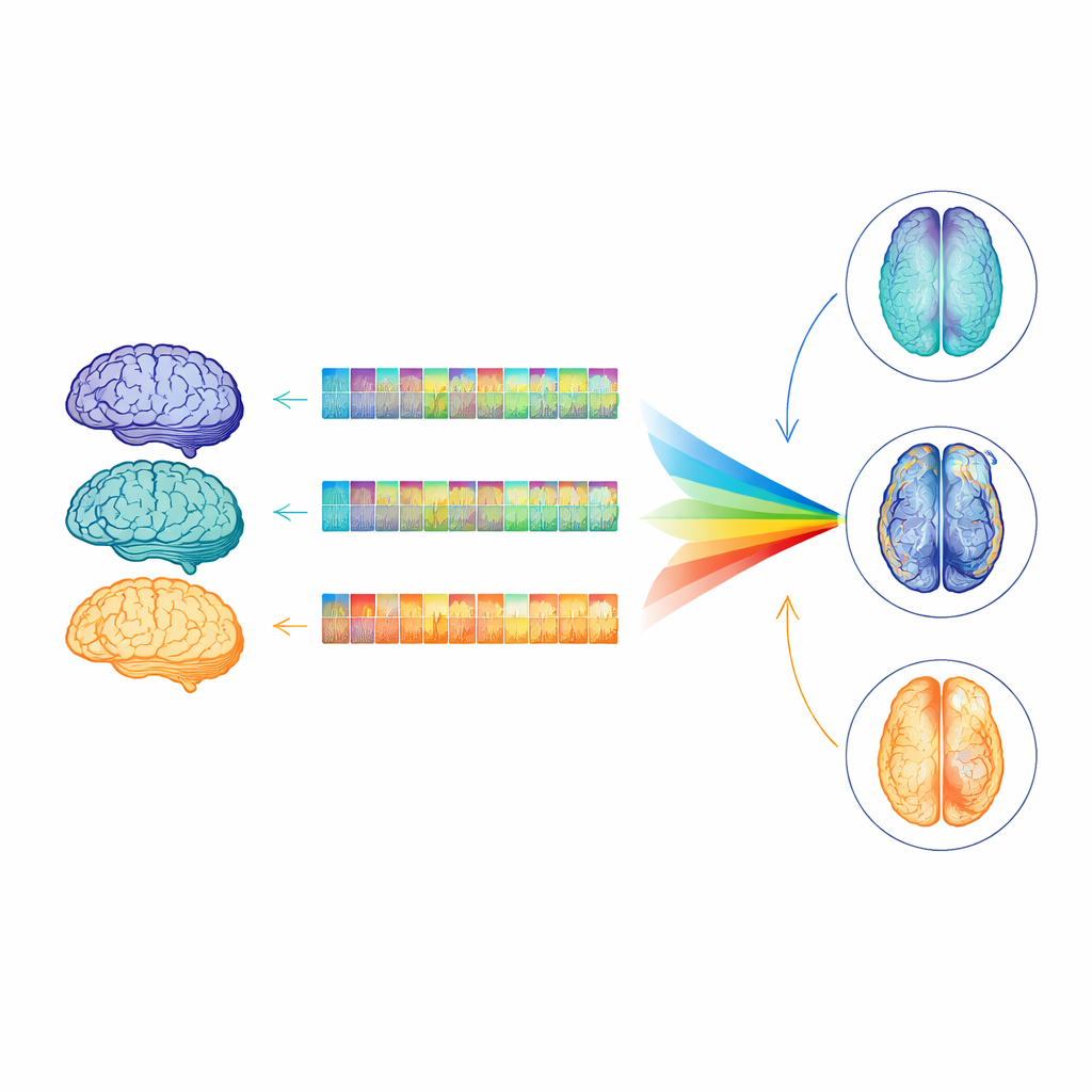
This careful design means that scientists can ask many “what if” questions about how fMRI is run and processed. Because the echoes differ in timing and image quality, they can be combined in smarter ways to boost the contrast between true brain activity and noise, or to estimate physical properties of brain tissue more precisely. The presence of phase data opens the door to advanced cleaning methods that isolate and subtract out physiology-related ripples in the signal, or to techniques that track fine shifts in the magnetic field over time. Side-by-side recordings on two scanners, with two repetition speeds and slightly different echo timings, allow direct tests of how these choices affect signal stability, brain coverage, and how strongly specific brain areas light up during the tasks.
Putting the data through its paces
To show that the dataset is robust, the authors ran a battery of quality checks. They quantified how much participants moved their heads, how stable the signal was over time, how well brain networks could be separated from noise, and how strongly key brain regions responded during the visual and oddball tasks. They found expected patterns: people moved more in the active tasks than at rest, faster scanning generally provided higher statistical power, and one scanner yielded slightly more stable signals than the other. Yet overall brain coverage remained remarkably consistent across scanners, task types, and timing settings, suggesting that the acquisition protocol is well-balanced and comparable. Group-level maps showed clear activation in visual and motor areas for the checkerboard task and more distributed responses for the oddball task.
What this means for future brain research
In plain terms, this work does not make a single headline-grabbing discovery about how the brain works; instead, it provides a carefully crafted test course on which many future “drivers” of brain analysis methods can train. By sharing a large, complex, and well-documented multi-echo fMRI dataset—with magnitude and phase data, several tasks, two scanners, and detailed heart and breathing recordings—the authors give the community a way to fairly compare new noise-removal tools, signal-combination strategies, and analysis pipelines. The ultimate payoff for the public is more trustworthy and informative brain imaging studies, whether they probe basic perception, monitor disease, or guide treatment.
Citation: Mikl, M., Ingrová, K., Gajdoš, M. et al. Complex multi-echo fMRI dataset: New strategies in processing of multi-echo data. Sci Data 13, 320 (2026). https://doi.org/10.1038/s41597-026-06694-6
Keywords: functional MRI, multi-echo imaging, brain mapping, neuroimaging methods, open data