Clear Sky Science · en
AIR-LEISH: A Dataset of Giemsa-Stained Microscopy Images for AI-based Leishmania amastigotes Detection
Why tiny parasites and smart cameras matter
Leishmaniasis is a parasite-borne disease that quietly affects millions of people, mainly in low‑income regions. Doctors and researchers still rely heavily on looking at stained blood and tissue smears under a microscope to spot the parasite inside immune cells—a painstaking process that can take hours and demands specialized training. This paper introduces AIR-LEISH, a freely available collection of microscope images designed to let computers learn to recognize these parasites automatically, opening the door to faster, cheaper, and more reliable tools for diagnosis and drug research.
From sandfly bites to hidden invaders
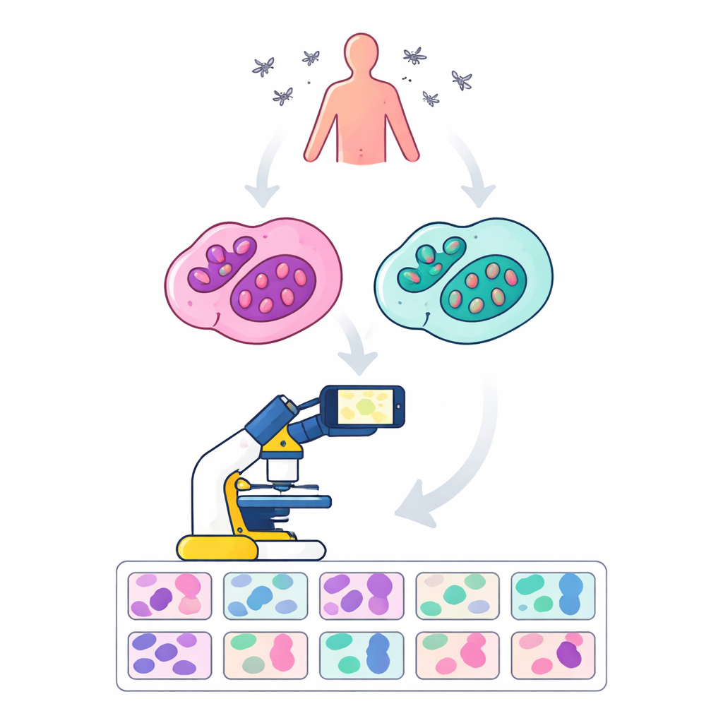
Leishmaniasis is spread by the bite of infected sandflies and can cause skin sores or life‑threatening infection of internal organs. The parasite lives and multiplies inside white blood cells called macrophages, hiding in a tiny round form known as an amastigote. To track how sick a patient is or how well a potential treatment works, researchers must count how many parasites are tucked inside these cells. Molecular lab tests can detect parasite DNA, but in many hospitals and research labs—especially in low‑resource settings—simple light microscopes remain the mainstay. Counting parasites by eye, however, is slow, tiring, and can vary from one observer to another.
Building a training set for artificial vision
Artificial intelligence has shown it can spot patterns in medical images that are too subtle or tedious for humans to process at scale. But to do this well, AI systems need thousands of carefully labeled examples. Until now, such image collections for leishmaniasis were scarce, incomplete, or difficult to access—especially for the clinically important amastigote stage inside cells. The authors created AIR-LEISH to fill this gap: 180 high‑resolution, Giemsa‑stained microscope images of infected human macrophages, captured with a regular smartphone mounted on a standard research microscope. Each picture shows cells from one of two infection setups, using different parasite species and host cell types, so that a wide range of realistic appearances is covered.
Turning raw pictures into trusted ground truth
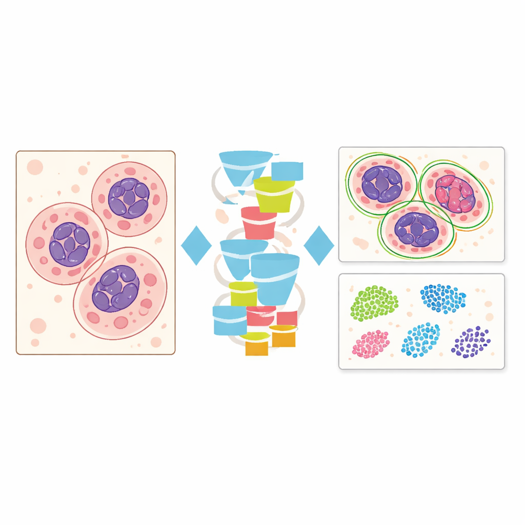
To make the images useful for computers, every cell and parasite had to be traced and labeled by hand. A parasitology expert first marked the outlines of individual macrophages, their nuclei, and the tiny amastigotes using a specialized annotation tool. An AI engineer then refined these markings pixel by pixel to ensure precise shapes and boundaries, including small or overlapping parasites. The team checked consistency between annotators and found a very high agreement, indicating that the labels can be trusted as ground truth. In total, the dataset includes 8,140 parasites, 1,511 host cells, and 1,731 nuclei, along with separate mask images that tell an algorithm exactly which pixels belong to which structure.
Putting AI models to the test
To show what AIR-LEISH can enable, the researchers trained two widely used image analysis systems. One, called U‑Net, is designed to color in each pixel according to whether it belongs to background, parasite, cell body, or nucleus. The other, YOLOv8, draws rectangular boxes around each object it detects and counts them. Despite the small size of the parasites and the limited number of images, both models did well at finding and separating parasites from their host cells, reaching high scores for both accuracy and reliability. The models even managed to spot a single infected cell among more than a hundred mostly clean cells, hinting at their potential to support very sensitive screening in the future.
Opening doors for better care and new cures
By releasing AIR-LEISH openly on the Zenodo platform, together with code and detailed documentation, the authors provide a practical foundation for many groups worldwide—especially those with limited resources—to build and compare AI tools for leishmaniasis. Because the images also include the host cells and their nuclei, the dataset can support broader studies of cell counting, infection levels, and even other pathogens that live inside similar immune cells. In simple terms, this work turns hours of expert microscope work into a reusable digital resource, helping speed up diagnostics, drug discovery, and ultimately the fight against a neglected but serious disease.
Citation: Oualha, R., Fekih-Romdhane, N., Driss, D. et al. AIR-LEISH: A Dataset of Giemsa-Stained Microscopy Images for AI-based Leishmania amastigotes Detection. Sci Data 13, 328 (2026). https://doi.org/10.1038/s41597-026-06676-8
Keywords: leishmaniasis, microscopy images, medical imaging AI, parasite detection, infectious disease diagnosis