Clear Sky Science · en
A Comprehensive X-ray Dataset for Pediatric Ulna and Radius Fractures Analysis
Why Broken Arms in Kids Matter
Broken forearms are a rite of passage for many active children, but spotting these fractures quickly and correctly on X-rays is not always easy. Doctors in busy emergency rooms may miss subtle breaks, especially in growing bones that look very different from those of adults. This article introduces a new open collection of children’s arm X-rays designed to help both doctors and computers get better at recognizing these injuries, potentially leading to faster, more reliable care. 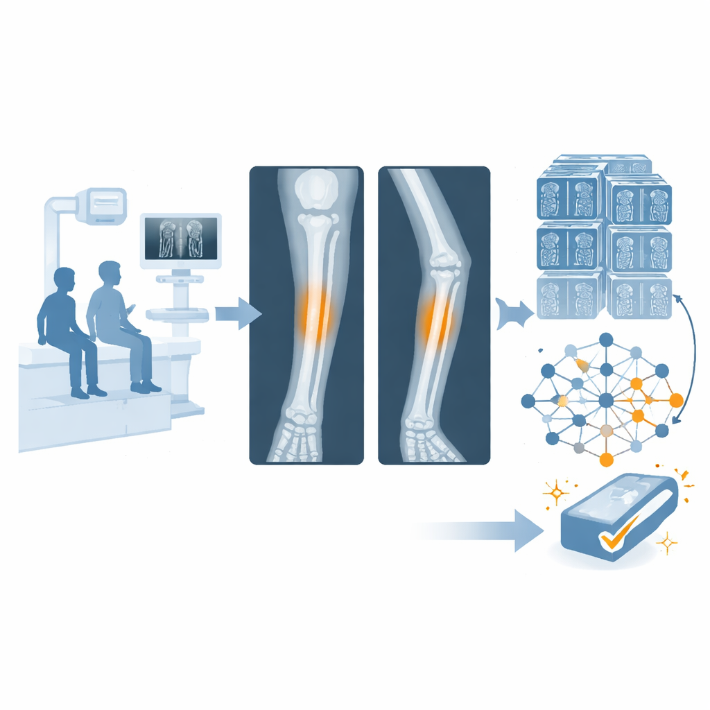
A New Library of Kids’ Arm X-Rays
The researchers created the Pediatric Ulna and Radius Fractures (PediURF) dataset, a large, public collection of more than 10,000 X-ray images of children’s forearm fractures. These images come from patients at a children’s hospital over more than a decade. Each X-ray has been stripped of names and other personal details to protect privacy. Importantly, every case includes two standard views of the forearm—one from the front and one from the side—because some fractures show up clearly in only one angle. Together, these paired views reflect how radiologists actually read images in everyday practice.
How the Images Are Carefully Labeled
To turn thousands of images into a useful scientific resource, experienced radiologists reviewed each case and assigned it to one of three locations along the forearm bones: near the elbow (proximal), in the middle (midshaft), or near the wrist (distal). These three regions matter because they are treated differently in the clinic and do not occur with the same frequency in real life. The dataset reveals that wrist-area fractures in children are by far the most common, mid-forearm breaks are less frequent, and elbow-area fractures are relatively rare but more complex. The images and these detailed labels together give researchers both visual variety and realistic statistics to train and test computer models.
How the Data Is Organized for Future Tools
The team divided the dataset into a training portion and a separate testing portion so that computer programs can be built and then fairly judged on images they have never seen before. Each child’s images stay entirely in one group to avoid overlap, and both the front and side views always travel together. Inside the folders, cases are sorted by fracture region and then by patient, with each patient folder holding exactly two X-ray files. This structure mirrors how data would appear in a hospital while still being simple enough for engineers to use in their code. The authors also share basic, non-identifying details such as age and sex in separate tables to enable more careful analysis.
A Test-Drive with a Smart Model
To show what can be done with PediURF, the researchers built a demonstration computer model called URFNet. This model takes in both X-ray views at once and runs each through a series of image-processing steps that gradually extract patterns, such as the outlines of the bones and the shape of a suspected break. A special "cross-attention" stage then allows information from the front view to influence how the side view is interpreted, and vice versa, mimicking how a human expert mentally compares the two angles. URFNet then decides whether the fracture is near the elbow, in the middle of the forearm, or near the wrist. In tests, it outperformed a wide range of well-known image-recognition systems, correctly classifying the vast majority of fractures even though some types were much rarer than others. 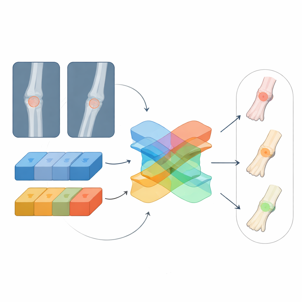
What This Means for Children’s Care
For parents and patients, the key takeaway is that this open X-ray library lays the groundwork for more reliable, faster computer assistance when a child shows up with a painful arm. Doctors, especially in busy or understaffed settings, could eventually use tools trained on PediURF to double-check their readings, highlight hard-to-see fractures, and prioritize urgent cases. While such systems will still need to be tested across many hospitals and refined to pinpoint exact fracture lines, this dataset marks an important step toward safer, more consistent care for some of the most common injuries in childhood.
Citation: Tang, S., Ou, L., Li, W. et al. A Comprehensive X-ray Dataset for Pediatric Ulna and Radius Fractures Analysis. Sci Data 13, 308 (2026). https://doi.org/10.1038/s41597-026-06666-w
Keywords: pediatric fractures, forearm X-rays, medical imaging AI, open medical datasets, deep learning radiology