Clear Sky Science · en
PMCanalSeg: A dataset for automatic segmentation of the pterygopalatine and mandibular canals from 3D CBCT images
Why Hidden Pathways in the Jaw Matter
When surgeons correct jaw deformities to improve a person’s bite or facial appearance, they work millimeters away from delicate nerves and blood vessels hidden inside bone. If these tiny channels are damaged, patients can suffer bleeding, numbness, or long‑lasting pain. This article describes PMCanalSeg, a newly released collection of 3D dental scans designed to help computers learn to spot two especially important bony canals in the upper and lower jaw, making these operations safer and more precise.
Delicate Tunnels Inside the Face
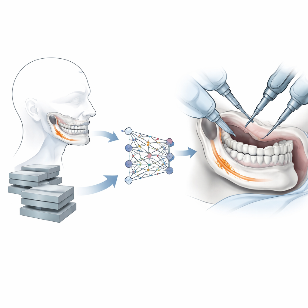
Inside our facial bones run narrow passageways that shelter nerves and vessels. Two of the most critical for jaw surgery are the mandibular canal, which carries the main nerve in the lower jaw, and the pterygopalatine canal, a smaller, more complex tunnel in the upper jaw. During orthognathic (jaw-correcting) surgery, doctors must cut and reposition bone while steering clear of these structures. Traditionally, surgeons or radiologists trace the canals slice by slice on cone beam CT (CBCT) scans, a 3D X‑ray method widely used in dentistry. This careful manual work is slow, requires deep expertise, and is vulnerable to human error.
Teaching Computers to See in 3D
In recent years, deep learning has transformed medical image analysis, allowing computers to learn how to outline organs and other structures automatically. However, these systems need many high‑quality, expertly labeled examples to reach clinical reliability. For mandibular canals, only a handful of public datasets exist, and they focus mainly on the lower jaw. A major blind spot has been the pterygopalatine canal in the upper jaw, which is harder to see and more variable from person to person. Without rich, open datasets spanning both canals, it is difficult to train robust algorithms or fairly compare different methods.
Building the PMCanalSeg Collection
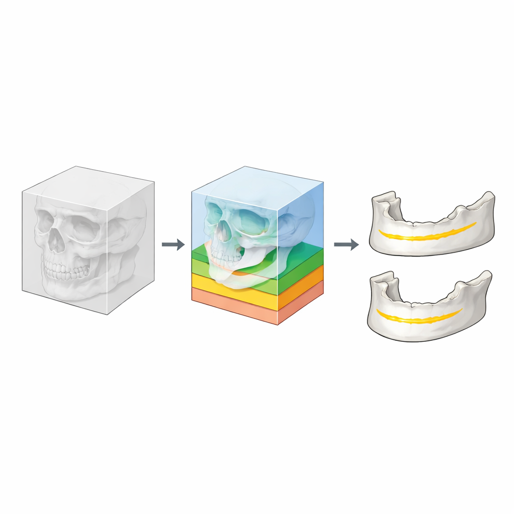
The authors address this gap by assembling PMCanalSeg, a curated set of CBCT scans from 191 patients treated at a dental hospital in China. All personal identifiers were removed under strict privacy rules, and only essentials such as age, sex, and scan date were kept. Each scan was converted from the original hospital format into a research‑friendly 3D file and processed to emphasize bone and remove unrelated structures like the spine. The skull was then digitally separated into upper and lower jaw regions so that algorithms can focus on the areas where the two canals run.
Expert Tracing and Careful Checking
To mark the canals accurately, four experienced oral surgeons worked in stages. Two specialists first traced the course of the pterygopalatine and mandibular canals on every 3D scan, defining which tiny 3D pixels belonged to each tunnel. Two additional surgeons then checked these markings layer by layer against the original images, correcting any discrepancies. For a sample of cases, the team measured how closely different experts agreed and found very high consistency, indicating that the labels are reliable. The final dataset is neatly organized by patient, with separate folders for upper jaw, lower jaw, and full skull volumes, making it straightforward for researchers to use.
How Well Do Machines Learn from It?
To test PMCanalSeg, the authors trained several leading 3D image‑segmentation networks and evaluated how closely their predictions matched expert labels. For the mandibular canal, modern transformer‑based models performed especially well, closely tracking the true nerve path. The pterygopalatine canal proved more challenging: its small size, complex shape, and the crowded anatomy of the upper jaw led to lower accuracy and more boundary errors. The team also compared results on PMCanalSeg with those on another widely used dataset for the lower jaw and discussed how differences in scan quality, labeling style, and anatomy coverage can alter reported performance.
What This Means for Patients and Research
For non‑specialists, the key message is that PMCanalSeg offers the first open collection of 3D jaw images with detailed markings for both a major lower‑jaw nerve canal and a previously neglected upper‑jaw canal. By making these data and supporting code freely available for non‑commercial use, the authors provide a strong foundation for developing and benchmarking computer tools that can automatically highlight these hidden pathways before surgery. As these tools improve, surgeons will be better able to plan cuts that avoid critical nerves and vessels, reducing complications and helping patients emerge from jaw surgery with safer, more predictable outcomes.
Citation: Li, G., Lu, Y., Wu, G. et al. PMCanalSeg: A dataset for automatic segmentation of the pterygopalatine and mandibular canals from 3D CBCT images. Sci Data 13, 312 (2026). https://doi.org/10.1038/s41597-026-06620-w
Keywords: cone beam CT, jaw surgery, medical image segmentation, dental imaging, deep learning dataset