Clear Sky Science · en
In-vivo optical properties spectra across five body locations on ten subjects using time-domain diffuse optics
Shining Light Deep Into the Body
Medical researchers increasingly use light, not X‑rays, to peer beneath the skin and track what is happening inside our bodies. But to turn light into a reliable diagnostic tool, scientists first need to know exactly how different tissues absorb and scatter it. This article presents a rich, openly available dataset that maps how light travels through living human tissue at several body sites, paving the way for safer, more precise optical tests and therapies.
Why Light Is a Powerful Medical Tool
Between red light and the near‑infrared, there is a “sweet spot” where light can penetrate centimeters into tissue without being completely absorbed. This range is already used in devices that monitor brain oxygen or guide laser treatments. However, most existing measurements of tissue “optical properties” come from pieces of tissue studied outside the body, from animals, or from small, fragmented experiments. That makes it hard to design new devices, compare studies, or account for natural differences between people. The authors set out to fill this gap with a standardized, in‑vivo human dataset that anyone can use.
How the Measurements Were Collected
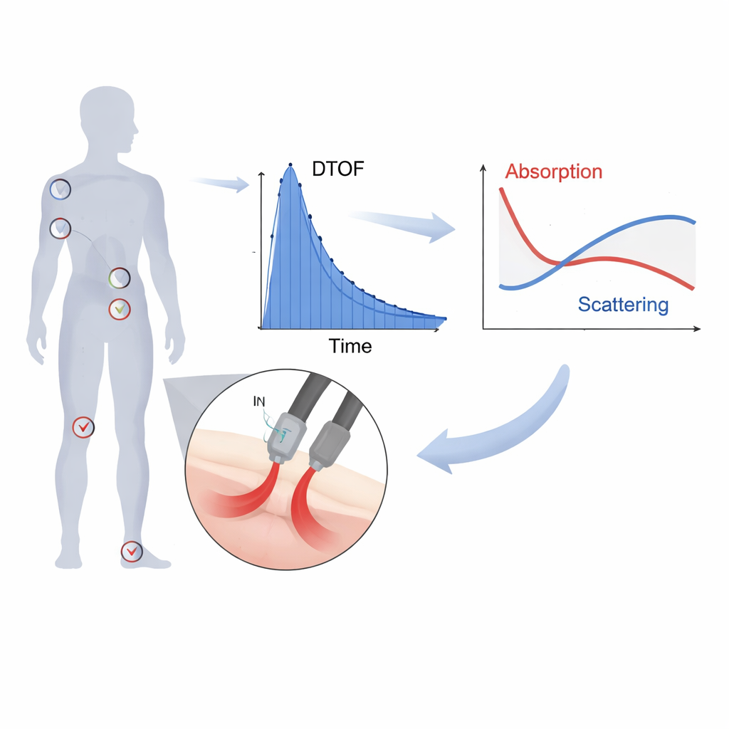
The team used a technique called time‑domain diffuse optical spectroscopy. They fired ultra‑short light pulses into the body through a small handheld probe and measured how long the scattered photons took to come back. The shape of this “time‑of‑flight” curve reveals how strongly tissue absorbs light and how much it scatters it. Ten healthy volunteers, differing in age, sex, skin tone, and body build, were measured at five locations: upper arm, forearm over the radius‑ulna bones, abdomen, forehead, and heel bone (calcaneus). For each site, light at 51 wavelengths from 610 to 1110 nanometers was recorded twice (with the probe repositioned) and three times per position, while ultrasound images were taken at the same spots to show the underlying anatomy.
Turning Photon Timings Into Tissue Maps
To translate the raw photon arrival times into something biomedically useful, the authors fitted each time‑of‑flight curve with a well‑tested physical model of light diffusion in scattering media. This allowed them to estimate two key numbers at every wavelength: how much light is lost to absorption, and how strongly it is scattered. The processing was done carefully to avoid noise and distortions, and the system was cross‑checked against liquid “phantoms” with known properties and against international performance benchmarks. The final dataset, hosted on Zenodo, includes the untouched raw files, metadata linking each file to subject and body site, example analysis outputs, and ready‑to‑use Python and MATLAB tools for reading and plotting the data.
What the Data Reveal About Real Bodies
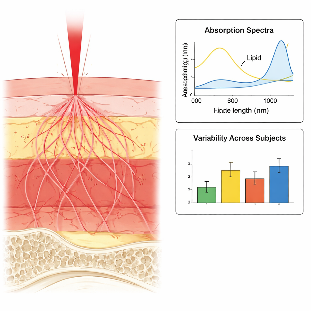
The resulting spectra show how water, fat, blood, and structural proteins each leave a distinct fingerprint in different parts of the body. For example, abdomen measurements in subjects with higher body mass index show stronger signals from fat at wavelengths where lipids absorb most, while leaner subjects show spectra dominated by water. Bone‑rich regions like the forearm and heel share subtle features likely linked to collagen in bone, and the forehead, which has little fat storage, is dominated by water and blood signatures. By comparing repeated measurements at the same spot to differences between people, the authors show that natural variation from person to person is much larger than the noise of the instrument itself, underlining how important it is to account for biological diversity when designing optical diagnostics.
A Foundation for Future Light‑Based Medicine
In everyday terms, this project is like building a detailed road map for how light travels through the body. Anyone designing a new optical scanner, testing a theory of how photons move in tissue, or training an artificial‑intelligence system to interpret optical signals can now start from accurate, openly shared human data rather than guesswork. By combining carefully validated measurements, ultrasound images, and transparent analysis tools, the dataset provides a common reference that should help accelerate the development of non‑invasive, light‑based methods to detect disease, monitor health, and guide therapy.
Citation: Damagatla, V., Karremans, S., Bossi, A. et al. In-vivo optical properties spectra across five body locations on ten subjects using time-domain diffuse optics. Sci Data 13, 261 (2026). https://doi.org/10.1038/s41597-026-06586-9
Keywords: tissue optics, near infrared light, noninvasive imaging, open biomedical data, photon migration