Clear Sky Science · en
PlaTiF: A pioneering dataset for orthopedic insights in AI-powered diagnosis of tibial plateau fractures
Why broken knees matter to more than doctors
Tibial plateau fractures are breaks in the upper part of the shinbone, right where it forms the knee joint. They may sound rare, but they can seriously affect walking, balance, and long‑term joint health, especially in older adults. Doctors rely on careful reading of X‑rays and scans to decide how to treat these injuries, yet this process is slow and not always consistent from one specialist to another. This article introduces a new, carefully prepared image collection designed to help computers learn to spot and classify these knee fractures, potentially making future care faster, fairer, and more reliable for patients.
The critical shelf that carries your weight
The tibial plateau is the flat, shelf‑like top of the shinbone that meets the thighbone to form the knee. It includes two rounded areas, called condyles, that cradle the cartilage and help the knee bend smoothly. When this region breaks—often after falls, traffic accidents, or sports injuries—the damage can extend beyond bone to nearby ligaments, meniscus, nerves, and blood vessels. Some fracture patterns are linked to hidden soft‑tissue injuries and may threaten the stability of the entire joint. Because the knee is central to standing and walking, getting the fracture type right is essential for planning surgery, predicting recovery, and avoiding arthritis later in life. 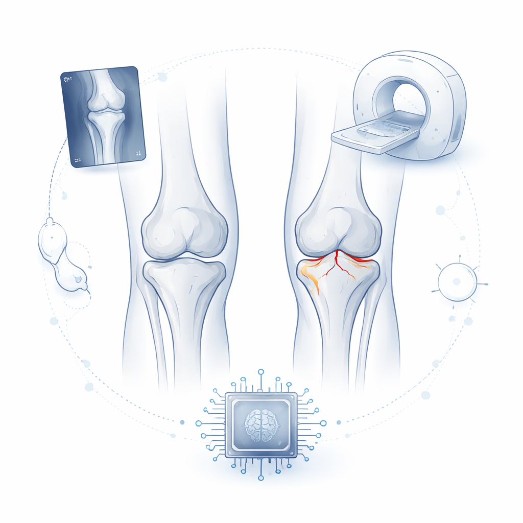
Why reading knee images is harder than it looks
Even for experienced orthopedic surgeons and radiologists, classifying these fractures can be tricky. A widely used system, known as the Schatzker classification, groups tibial plateau fractures into six main types based on where the bone is broken and how badly it is crushed or shifted. Traditionally, doctors use standard front‑view X‑rays of the knee, sometimes supported by CT scans, to decide which type they are dealing with. However, X‑rays can be blurred by overlapping bones, low contrast, or patient positioning, and CT scans are expensive and expose patients to higher radiation. As a result, different doctors may disagree about the same image, and creating computer tools that mimic expert judgment has been held back by a shortage of well‑labeled examples.
A new open collection of real‑world knee images
The researchers behind this work created PlaTiF, the first open‑access dataset focused specifically on tibial plateau fractures for use in artificial intelligence (AI) research. It gathers 421 X‑ray images from 186 patients, along with supporting CT slices for each case. Every knee was evaluated by multiple orthopedic experts, who assigned a Schatzker fracture type—or recorded that the tibial plateau was normal—after reaching consensus on difficult cases. The final dataset includes a broad mix of patients, with a mean age of about 46 years and a full spread of fracture types, from simple cracks to complex, multi‑piece breaks. For each image, the team also recorded demographic details and which side of the body was injured, packaging everything in a structured format that is easy for researchers to load into analysis software. 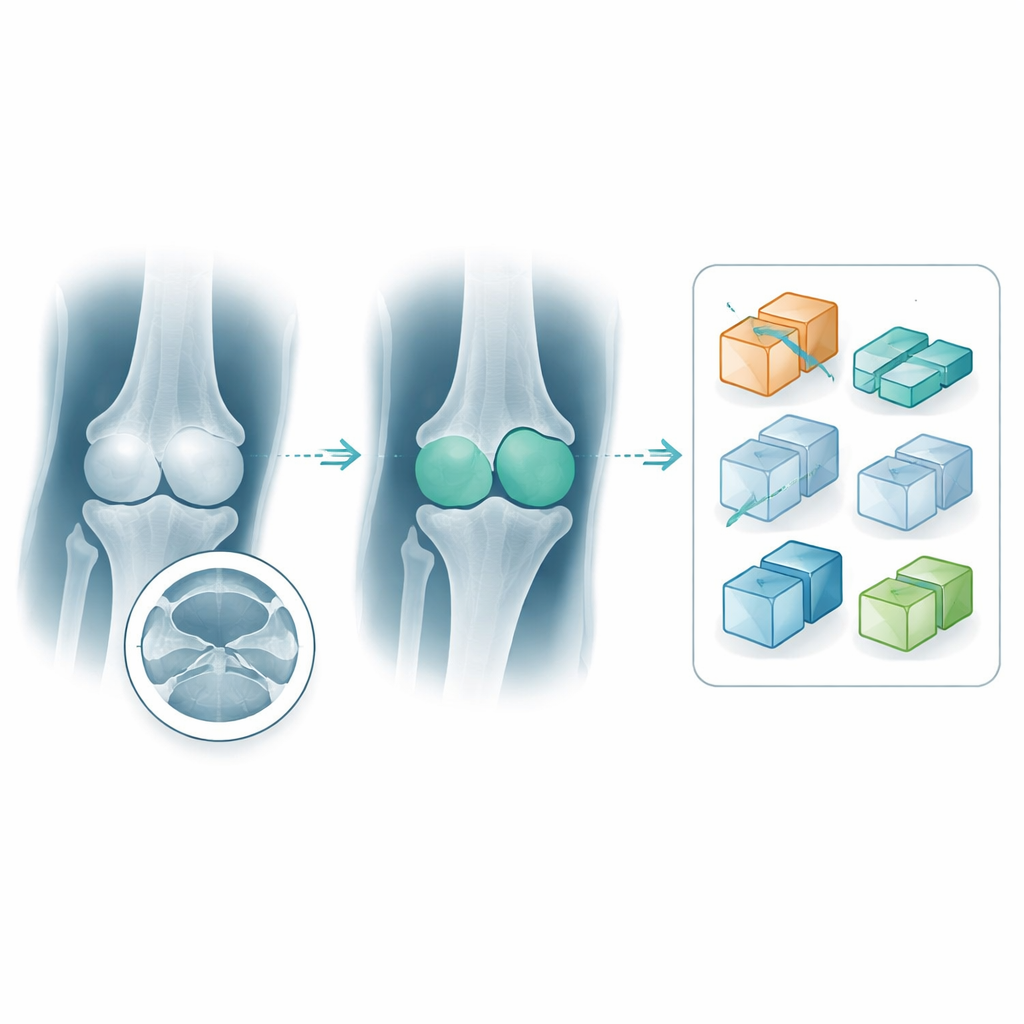
Teaching computers to see the bone more clearly
Beyond simple labels, PlaTiF also includes detailed outlines separating the tibia from nearby bones and background tissue. To create these “masks,” specialists used an interactive image‑processing tool: an algorithm first guessed the bone area, and then experts refined the edges by hand and applied shape‑cleaning steps. The result is a set of binary masks that cleanly highlight the tibia in each X‑ray. These masks are crucial for training AI systems not only to say whether a fracture is present, but also to focus on the right anatomical region and learn how different fracture patterns change the shape and surface of the bone. The authors envision researchers using these data to build and compare machine‑learning models, generate realistic synthetic examples to balance rare fracture types, and eventually support clinical decisions such as surgical planning.
Ensuring quality today, planning for better tools tomorrow
To make sure PlaTiF can be trusted as a training ground for AI, the team followed strict quality‑control procedures. Multiple experts independently reviewed every fracture label and bone outline, resolving disagreements through discussion until they reached full agreement. All data were anonymized and released under an open license, so scientists worldwide can download, test, and improve their methods. While the first version includes only front‑view X‑rays, the authors plan to add side views and full CT scans in the future, which will better capture the three‑dimensional shape of fractures. For patients, the long‑term promise is that AI systems built on resources like PlaTiF could help doctors detect subtle injuries earlier, choose treatments more precisely, and improve the chances of a stable, pain‑free knee after a serious injury.
Citation: Kazemi, A., Same, K., Zamanirad, A. et al. PlaTiF: A pioneering dataset for orthopedic insights in AI-powered diagnosis of tibial plateau fractures. Sci Data 13, 240 (2026). https://doi.org/10.1038/s41597-026-06560-5
Keywords: tibial plateau fracture, knee X-ray, medical imaging dataset, orthopedic AI, fracture classification