Clear Sky Science · en
PhysioMio: bilateral and longitudinal HD-sEMG dataset of 16 hand gestures from 48 stroke patients
Why This Matters for Life After Stroke
After a stroke, simple actions like buttoning a shirt or holding a cup can become daily challenges. Doctors and therapists work hard to help people regain hand and arm function, but they often have to rely on what they see and what patients report. The PhysioMio project changes this by providing a large, open scientific dataset that turns invisible muscle activity into precise, measurable information. This can help researchers build smarter rehabilitation tools, more responsive assistive devices, and ultimately more personalized therapy plans for stroke survivors.
Listening to Muscles Through the Skin
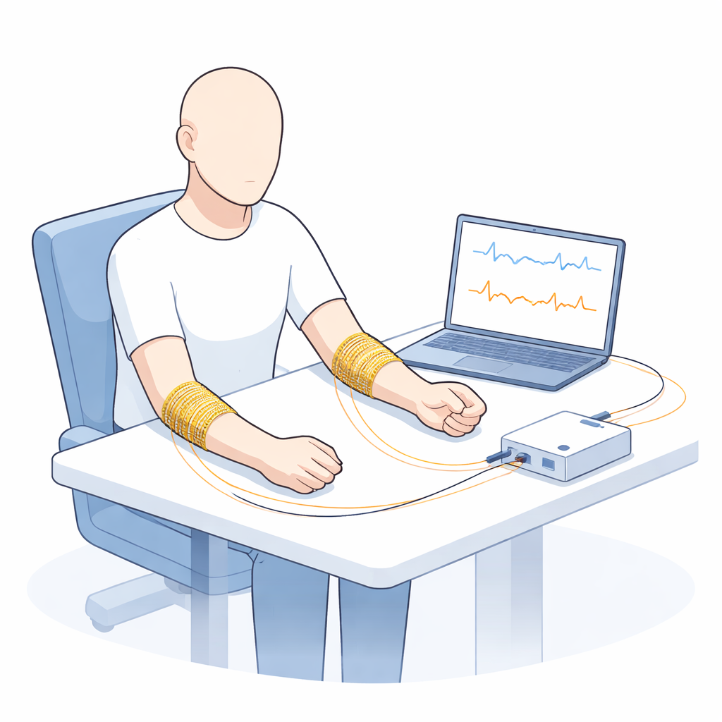
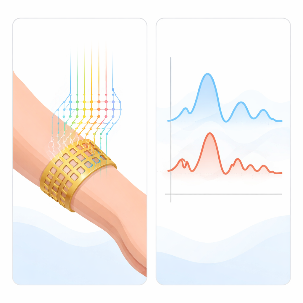
Every voluntary movement starts with tiny electrical signals in our muscles. PhysioMio uses a technique called surface electromyography, which works like a very sensitive stethoscope for muscles placed on the skin. Instead of just one or two sensors, the team wrapped a band of 64 small, dry metal contacts around the forearm. This high-density setup captures a detailed map of how different muscle groups fire when a person tries to move their hand and fingers. Because the sensors sit on the skin, the method is non-invasive and safe to repeat many times during recovery.
A Large Group of Real Stroke Patients
The dataset comes from 48 people who had a stroke and were undergoing rehabilitation. They varied widely in age, body size, and time since stroke, reflecting the diversity seen in real clinics. For each patient, the researchers recorded up to 16 hand and wrist gestures, including resting, several types of grasp (such as pinching and holding a ball), and wrist bending in different directions. Importantly, they recorded both the healthy arm and the affected arm, and they followed patients over multiple sessions during their stay in rehabilitation. That means the data not only shows how different the impaired arm is from the healthy one, but also how muscle activity can change over time as people recover.
How the Measurements Were Collected
During each session, a flexible electrode band was carefully placed around the forearm at a fixed position, disinfected beforehand to ensure good contact and hygiene. Patients sat comfortably while a trained investigator demonstrated each gesture and gave clear instructions. Once the patient reached the best possible position for a gesture, a foot-operated switch marked the time interval of interest, so the computer knew exactly when the movement happened. Each recording lasted about 10 to 15 minutes and included all 16 gestures. Later, the central four seconds of muscle activity for every gesture were cut out and saved in a standard, efficient file format. Alongside the muscle signals, the dataset includes information such as age, gender, which arm was affected, and how many days had passed since the stroke, allowing researchers to link muscle patterns to the course of recovery.
Making Sure the Signals Are Trustworthy
To be useful, such a dataset must have clean, reliable measurements. The team followed strict procedures before, during, and after each recording. They checked signal quality with test contractions, reduced electrical interference from the power grid, and stored all data securely and anonymously. Afterward, they visually inspected every recording and removed sessions where many electrodes failed or the signal was too noisy. They then used mathematical checks to confirm quality, such as comparing the strength of muscle activity during movement to rest and analyzing how the signals were distributed and spread across different frequencies. Finally, they trained a simple computer model that could tell, with high accuracy, whether a recording came from a healthy or impaired arm, which further confirmed that the dataset captures real, meaningful differences in muscle function.
What This Opens Up for Future Care
In everyday terms, the PhysioMio dataset is a detailed logbook of how damaged and healthy muscles behave as people try to move their hands after a stroke. Because it is publicly available and well documented, scientists and engineers around the world can use it to design better movement detectors, smarter rehabilitation robots, and more objective tests of hand function. Over time, such tools could help therapists spot early signs of improvement or problems, and tailor exercises to each individual. For stroke survivors, that could mean more efficient therapy, better support for daily activities, and a clearer path from hospital care back to independent living.
Citation: Ilg, J., Oldemeier, A.C.R., Fieweger, M. et al. PhysioMio: bilateral and longitudinal HD-sEMG dataset of 16 hand gestures from 48 stroke patients. Sci Data 13, 19 (2026). https://doi.org/10.1038/s41597-026-06557-0
Keywords: stroke rehabilitation, electromyography, hand function, neuromuscular recovery, assistive technology