Clear Sky Science · en
Base barrier cells provide compartmentalization of choroid plexus, brain and CSF
Hidden Walls Inside the Brain
The brain floats in a clear liquid called cerebrospinal fluid, or CSF, and is guarded by several biological “walls” that control what can get in or out. This study reveals a previously unknown wall at the root of the choroid plexus—a frond-like tissue that makes CSF inside the brain’s ventricles. Understanding this hidden barrier helps explain how the brain keeps blood-borne substances and immune cells at bay, and what may go wrong during inflammation or disease.
A Gate Between Brain, Blood, and Fluid
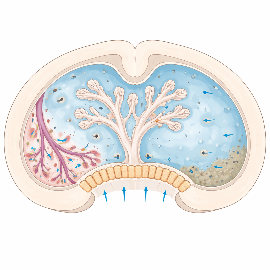
The choroid plexus sits in fluid-filled cavities and produces much of the CSF that bathes the brain and spinal cord. Scientists already knew that an epithelial cell layer in the choroid plexus acts as a blood–CSF barrier. But at the spots where this tissue attaches to the brain surface, there was an anatomical puzzle: blood vessels running through the leaky interior of the choroid plexus seemed to touch spaces filled with CSF and the brain’s outer layers. Without an additional barrier, substances from blood and stromal tissue could spill directly into the CSF and surrounding brain. The authors set out to see whether a specialized cell population at this attachment zone quietly performs this protective role.
Finding a Special Cell Type
Using single-cell RNA sequencing in mice, the researchers cataloged thousands of individual cells from the choroid plexus and nearby coverings of the brain. They discovered two distinct fibroblast-like cell types: one spread throughout the inner core (stroma) of the choroid plexus, and another concentrated only at its base, where it fastens to the brain and lies next to CSF-filled spaces. These “base” cells carried a genetic fingerprint similar to known barrier cells in the meninges—the membranes surrounding the brain—suggesting they might act as a seal. Developmental tracing experiments showed that these cells arise early in embryonic life from the same mesenchymal tissue that forms the meninges and then persist with a stable identity into old age.
How the Base Cells Build a Seal
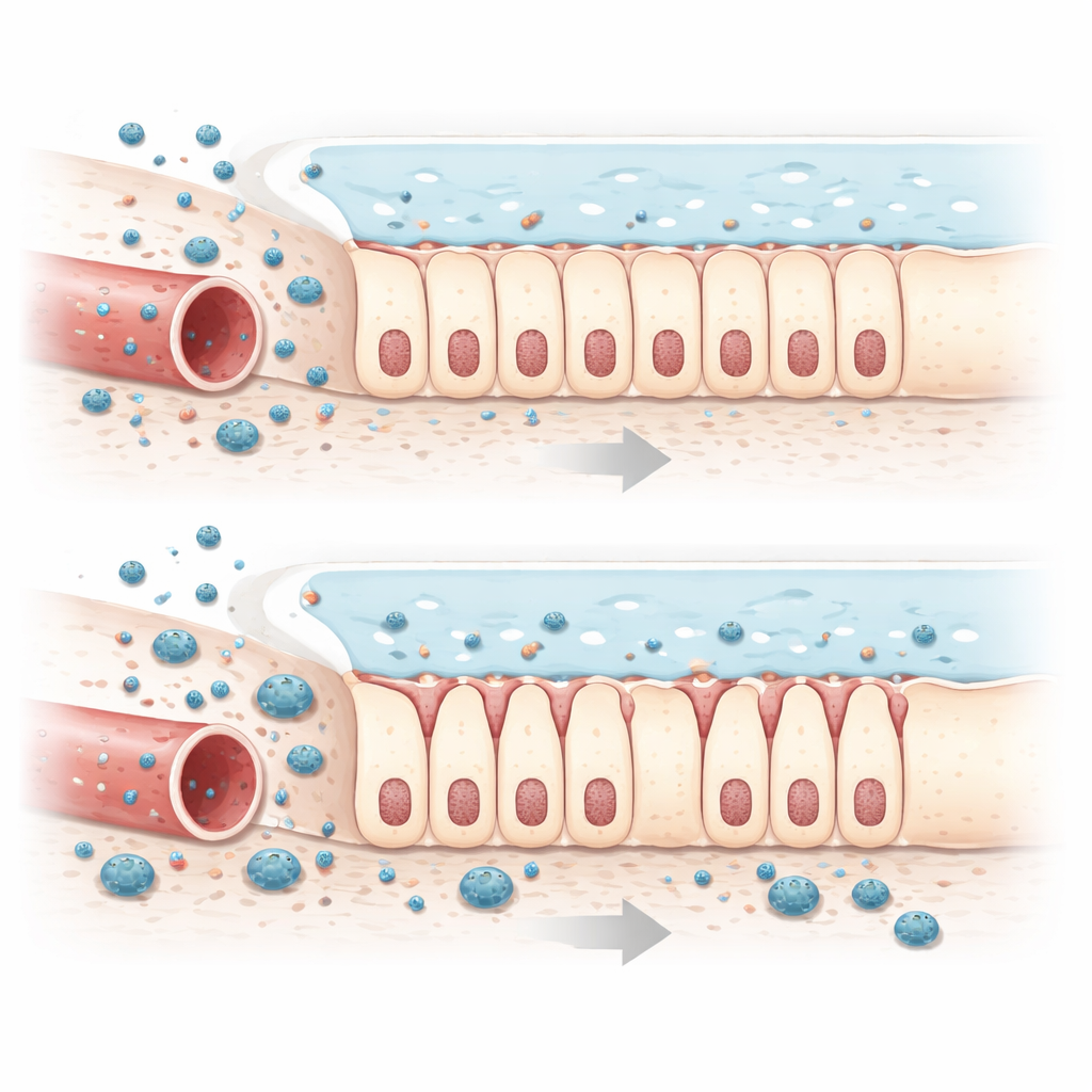
Microscopy at multiple scales, from confocal imaging to three-dimensional electron microscopy, revealed that the base cells pack together into a dense cluster encasing arterioles and venules as they enter the choroid plexus. The cells interlock through tight and adherens junctions—specialized contact sites that fuse neighboring cell membranes. Rather than laying down thick collagen or other scaffold material, these cells behave more like living caulking, forming a continuous plug between three compartments: the leaky interior of the choroid plexus, the brain tissue, and the CSF in the ventricles and subarachnoid space. When small tracer molecules were injected into the bloodstream, they could escape from fenestrated capillaries into the choroid plexus stroma but stopped abruptly at this plug. When tracers were placed directly into CSF, they washed over the brain and choroid plexus surface yet again failed to cross into the stromal side. Together, these tests showed that the base cells function as an effective two-way barrier.
When Inflammation Breaks the Seal
The team then asked what happens during systemic inflammation, a whole-body immune response that is known to weaken other brain barriers. After giving mice a bacterial component that triggers inflammation, the researchers saw reduced expression of key junction proteins in the base cells and more diffuse, open contacts between them under the electron microscope. Tracer molecules that previously stopped at the base now leaked into and across this region. Immune cells called monocytes, normally confined to the inner choroid plexus tissue, accumulated at the base and were observed on the CSF side of the cell layer, indicating that this site can become a gateway for immune entry when the barrier is stressed.
A Conserved Shield With Broad Relevance
Finally, by comparing mouse data to single-nucleus sequencing of human choroid plexus, and by staining human postmortem tissue, the authors identified an analogous cell population in people. These human cells sit at the choroid plexus base, show the same characteristic markers, and form a honeycomb-like junction pattern suggestive of a seal. The findings establish “base barrier cells” as a conserved, lifelong barrier population that compartmentalizes the choroid plexus, brain tissue, and CSF. For non-specialists, the key message is that there is an additional, previously unrecognized wall inside the fluid spaces of the brain. When intact, it helps keep blood-derived molecules and immune cells from freely mixing with the brain’s delicate environment; when weakened by inflammation, it may open a new route for harmful substances and cells to enter, with potential implications for infections, autoimmune diseases, and other neurological disorders.
Citation: Verhaege, D., De Nolf, C., Van Acker, L. et al. Base barrier cells provide compartmentalization of choroid plexus, brain and CSF. Nat Neurosci 29, 551–566 (2026). https://doi.org/10.1038/s41593-025-02188-7
Keywords: choroid plexus, brain barriers, cerebrospinal fluid, neuroinflammation, meningeal fibroblasts