Clear Sky Science · en
Functional network comparative area and topography analysis (FUNCATA) in non-affective psychosis: a replication study
Why Brain Wiring in Psychosis Matters
Psychotic disorders like schizophrenia are often described in terms of unusual thoughts or perceptions, but underneath these symptoms lies a complex story about how brain networks are organized. This study asks a deceptively simple question: are the main communication systems of the brain physically laid out differently in people with early psychosis? By zooming in on the size and location of large-scale brain networks in living people, the researchers hope to find reliable brain-based markers that could one day help with earlier detection and more personalized treatment.
Looking at the Resting Brain
The researchers used resting-state functional MRI, which captures slow, spontaneous activity patterns while people lie quietly in the scanner. These patterns reveal how distant brain regions naturally coordinate, forming large networks that support everyday functions such as attention, movement, and inward-focused thought. Instead of relying on a one-size-fits-all brain map, the team used an approach called FUNCATA to carve out each person’s networks individually. This method measures how big each network is on the cortex and how closely its borders match a reference map built from over a thousand healthy young adults.
Comparing Healthy Brains and Early Psychosis
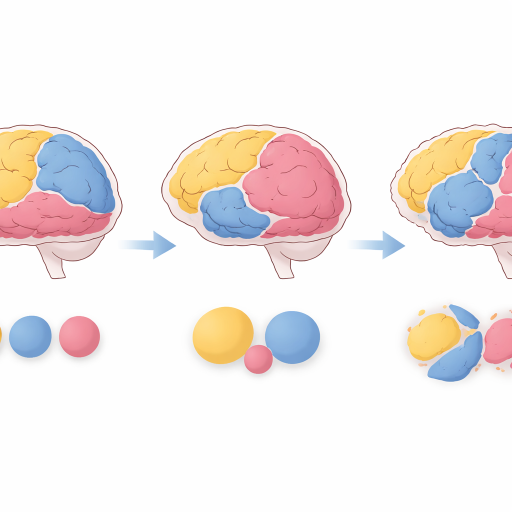
The study examined 86 young adults with non-affective psychosis and 57 healthy peers. It focused on ten major networks, including those involved in paying attention to the outside world, daydreaming and self-reflection, and moving the body. Compared with healthy participants, the psychosis group had a larger dorsal attention network, which helps focus on relevant sights and sounds, and a larger default mode network, which supports internal mentation such as mind-wandering. In contrast, a sensorimotor-body network, tied to basic movement and bodily sensations, tended to be smaller in psychosis. These size differences were moderate in magnitude and remained even after accounting for age and head motion during scanning.
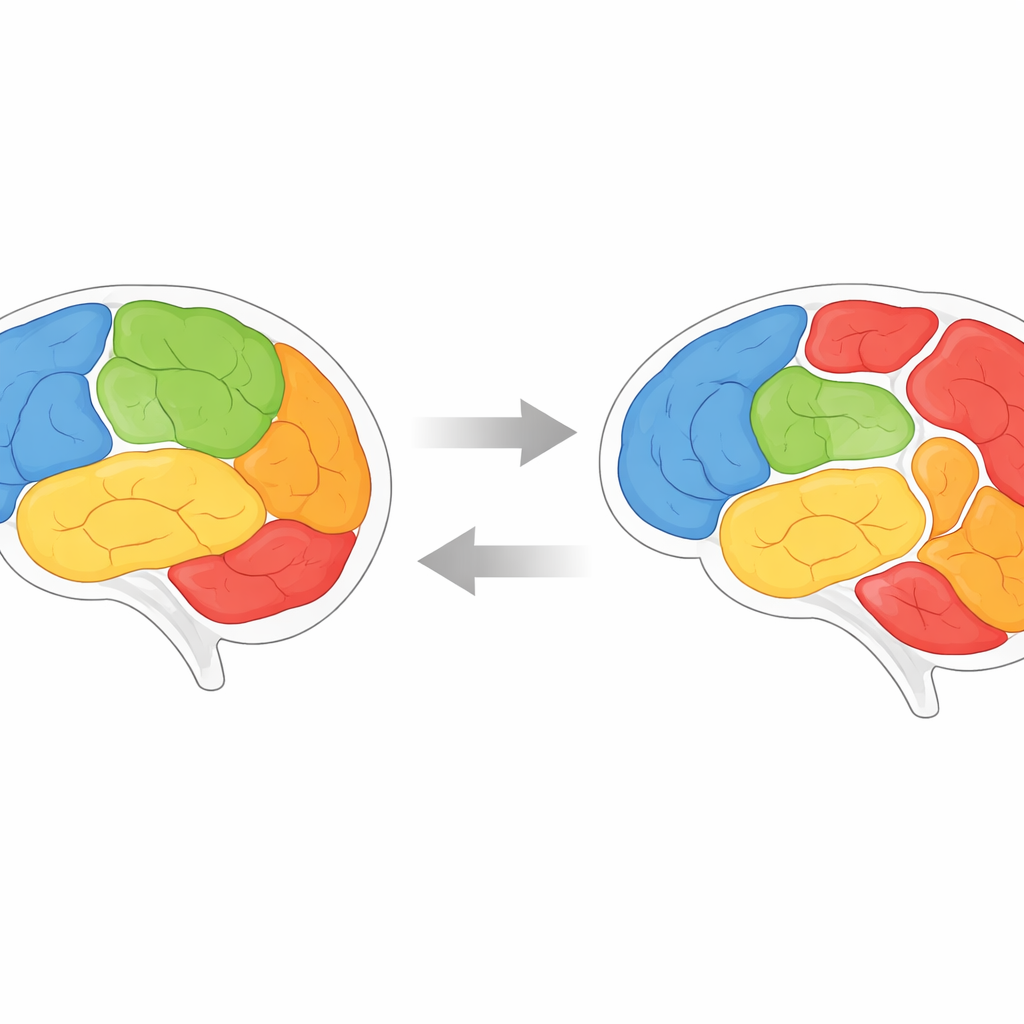
When Brain Maps Shift Their Borders
Size alone did not tell the whole story. The team also asked whether each network sat where it “should” according to the reference map. To do this, they introduced a Topographic Abnormality Index, which measures how much of a person’s network spills beyond its typical territory. People with psychosis showed greater topographic abnormality in the default mode, dorsal attention, and cingulo-opercular networks, the last of which is important for maintaining goals and monitoring performance. In some regions, areas that would usually be assigned to one network in healthy brains were more often claimed by a different network in psychosis, suggesting a subtle tug-of-war over cortical real estate.
Different Brain Types Within Psychosis
Psychotic disorders are clinically diverse, and the brain data reflected that diversity. Using patterns of network size, the researchers identified three “biotypes” within the psychosis group. One subgroup had enlarged attention and language networks and tended to show higher medication exposure and more classic schizophrenia diagnoses. Another subgroup had network profiles close to healthy controls but more prominent mood symptoms. A third showed enlarged default mode and attention networks along with smaller frontoparietal and sensorimotor-body networks and more negative symptoms. Across these biotypes, greater deviations in network layout often tracked with worse thinking skills, such as working memory, vocabulary, and emotion recognition.
What This Means for People With Psychosis
Taken together, the findings suggest that in early psychosis, key brain networks are not only communicating differently but are literally stretched, shrunk, or shifted on the brain’s surface. The consistent enlargement and misalignment of the attention and default mode networks hint that these systems could serve as early brain markers of psychosis risk, while changes in other networks may reflect illness stage or symptom patterns. Although no single measure is accurate enough to diagnose a person, combining individualized network maps with clinical information could eventually help predict who is at highest risk, who might respond best to certain treatments, and how illness unfolds over time.
Citation: Mamah, D., Chen, S., Harms, M.P. et al. Functional network comparative area and topography analysis (FUNCATA) in non-affective psychosis: a replication study. Schizophr 12, 32 (2026). https://doi.org/10.1038/s41537-026-00736-z
Keywords: schizophrenia, brain networks, resting-state fMRI, early psychosis, functional connectivity