Clear Sky Science · en
In-vivo histology of Parkinson’s disease using quantitative multiparametric mapping
Why Looking Inside the Living Brain Matters
Parkinson’s disease is usually recognized by its outward signs—tremor, stiffness, and slowed movement—but the real story unfolds deep inside the brain. This study shows how a new type of MRI can act like a virtual microscope, revealing tiny changes in brain tissue in people living with Parkinson’s. By detecting these changes early and across the whole brain, doctors may one day track disease progression more precisely and tailor treatments to each individual.
A Closer Look at Parkinson’s Beyond Movement
Parkinson’s is often described as a disorder of one small region, the substantia nigra, where dopamine-producing cells die. Yet patients also experience problems with thinking, mood, and motivation, hinting that the disease extends far beyond this single area. The authors set out to map subtle shifts in brain structure across both gray matter (the brain’s “processing units”) and white matter (the “wiring” that connects them). Rather than focusing only on late-stage damage, they asked whether these changes could already be seen in people with mostly mild to moderate symptoms.
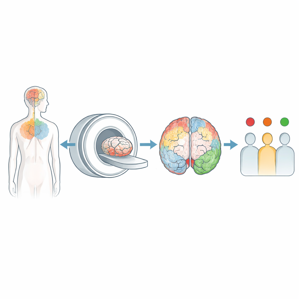
A Virtual Biopsy Using Advanced MRI
To peer into the living brain with greater detail, the team used a technique called multiparametric mapping, a form of quantitative MRI. Unlike conventional scans that give mostly anatomical pictures, this approach measures several physical properties of tissue that are linked to biology: how quickly signals relax, how much water is present, and how strongly molecules interact with surrounding structures. These measures are sensitive to myelin (the insulation around nerve fibers), iron deposits, and overall cell and water content—features that typically can only be examined under a microscope after death. In this study, 31 people with Parkinson’s and 68 similar healthy volunteers underwent a roughly half-hour scan that produced whole-brain maps of these properties.
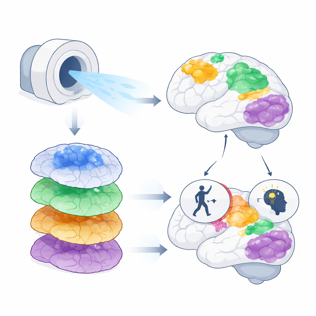
Hidden Brain Changes Tied to Movement and Memory
The maps revealed widespread differences between people with Parkinson’s and healthy controls, especially in the frontal lobes, the cingulate cortex, parietal areas, and the cerebellum. In several regions important for movement planning and control—such as the supplementary motor area and superior frontal gyrus—the tissue signatures suggested a mix of iron buildup, myelin disruption, and other forms of remodeling. Some of these changes lined up with how patients were doing clinically. Lower values in a frontal region called the superior frontal gyrus were linked to worse motor scores, meaning more severe movement problems. In parietal regions that support spatial awareness and high-level thinking, altered tissue properties were associated with lower scores on a brief cognitive test, indicating more pronounced thinking difficulties.
Patterns Across Multiple Brain Systems
Intriguingly, many of the affected gray-matter areas had corresponding changes in nearby white-matter tracts, suggesting that Parkinson’s disrupts both the local processing hubs and the connections that join them. Measures linked to iron content often changed together with those related to myelin and water, pointing to a complex blend of inflammation, loss of nerve fibers, and possible attempts at repair. At the same time, the researchers did not see clear differences in some of the classic deep-brain nuclei, including the substantia nigra, in this mostly early- to mid-stage group. This supports the idea that certain hallmark changes, such as heavy iron accumulation in these nuclei, may emerge later in the disease or evolve in a more gradual, stage-dependent fashion.
What This Means for People with Parkinson’s
For patients and clinicians, the message is cautiously hopeful. This work shows that a single, non-invasive MRI protocol can detect biologically meaningful microstructural changes across the entire brain, and that some of these changes track with how people move and think. While more research is needed—especially long-term studies and wider clinical use—multiparametric mapping could become a powerful tool for monitoring disease progression, testing new therapies, and ultimately personalizing care. Instead of waiting for symptoms to worsen or for major shrinkage on standard scans, doctors may be able to watch the disease at work in real time and intervene more intelligently.
Citation: Pokotylo, M.M., Göttlich, M., Schmidt, L. et al. In-vivo histology of Parkinson’s disease using quantitative multiparametric mapping. npj Parkinsons Dis. 12, 82 (2026). https://doi.org/10.1038/s41531-026-01329-4
Keywords: Parkinson’s disease, brain MRI, microstructural imaging, neurodegeneration, personalized neurology