Clear Sky Science · en
Neuromelanin imaging outperforms free water imaging in diagnosing early Parkinson’s disease: a comparative MRI study
Why catching Parkinson’s early matters
Most people think of Parkinson’s disease as a condition that reveals itself only when shaking, stiffness, and slowness become obvious. In reality, by the time those outward signs appear, many of the brain cells that control movement have already been lost. Doctors urgently need reliable, easy-to-use tools that can spot Parkinson’s earlier and more accurately, so that treatment and research on disease-slowing therapies can begin before too much damage is done.
Looking inside the movement center of the brain
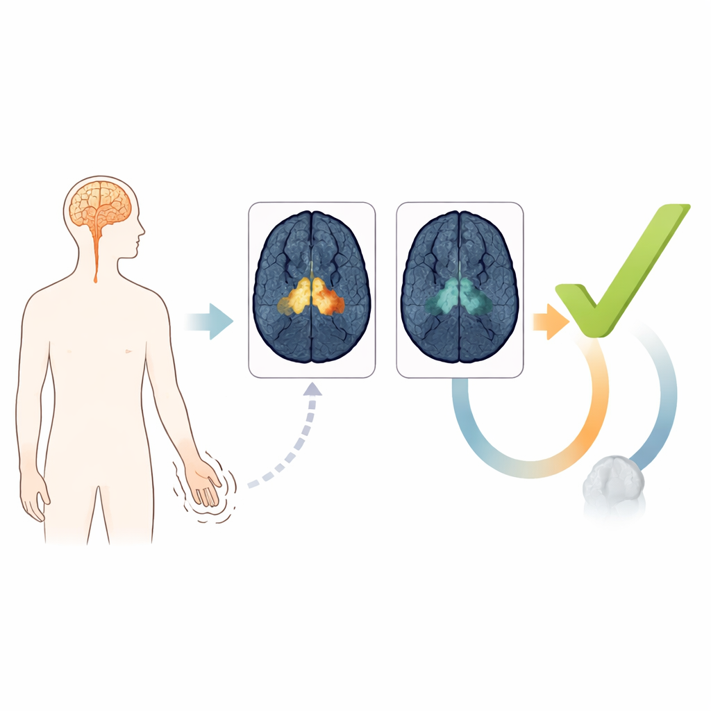
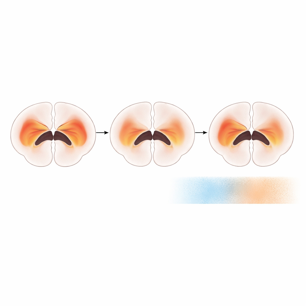
In Parkinson’s disease, a small deep-brain region called the substantia nigra slowly loses nerve cells that make dopamine, a chemical crucial for smooth movement. These cells contain a dark pigment known as neuromelanin, which can be seen on specialized MRI scans. Another MRI approach, called free-water imaging, looks instead at how water moves in brain tissue and can reflect swelling, inflammation, or loss of structure. The study asked a simple but important question: when it comes to spotting early Parkinson’s, which of these two MRI signals tells us more?
Comparing two MRI “biomarkers” head to head
The researchers analyzed brain scans from 247 people with early Parkinson’s and 78 comparison patients who had symptoms such as tremor or dizziness but were ultimately found not to have Parkinson’s. Everyone underwent standard MRI plus two advanced add-ons: neuromelanin-sensitive imaging and diffusion imaging for free-water measurement. The team focused on the substantia nigra and divided it into three functional zones related to movement, emotion, and thinking. Sophisticated automated software measured neuromelanin-rich volume and free water in each zone, greatly reducing human bias and making it feasible to process large numbers of scans in a consistent way.
Neuromelanin loss stands out; water changes do not
Across all parts of the substantia nigra, people with early Parkinson’s showed a clear loss of neuromelanin-rich tissue compared with controls, especially in the movement-related zone. In contrast, free-water levels were surprisingly similar between the two groups on the same scanner and analysis pipeline. When the team tested how well each measure could distinguish early Parkinson’s from non-Parkinsonian conditions, neuromelanin-based measures consistently outperformed free-water measures. A combined model that blended information from all three neuromelanin zones worked best, correctly classifying patients more often than any single region alone.
Holding up in the earliest and independent cases
The advantage of neuromelanin imaging held even when the analysis was restricted to patients who had experienced symptoms for less than two years, a window especially relevant for early diagnosis and for recruiting people into clinical trials. The researchers then repeated the comparison in an independent group from another hospital, using different MRI machines but similar techniques. Once again, neuromelanin volume was lower in early Parkinson’s and better at separating patients from controls than free water. The study also found that neuromelanin loss tracked with how long someone had been living with Parkinson’s, while free-water changes were more strongly influenced by age and sex than by disease itself.
What this means for patients and future care
For someone worried about Parkinson’s, these findings point toward neuromelanin MRI as a promising tool that looks directly at the nerve cells most affected by the disease, rather than at secondary changes in the surrounding tissue. While this technique is not yet part of routine diagnosis and still requires careful standardization, the results suggest it could help doctors recognize Parkinson’s earlier and with more confidence, select the right participants for trials of disease-slowing drugs, and eventually track how well those treatments protect the brain. Free-water imaging may still prove useful later in the illness, but in the critical early phase, neuromelanin imaging appears to give the clearest picture of what is going wrong inside the movement center of the brain.
Citation: Roh, Y.H., Youn, J., Kim, SY. et al. Neuromelanin imaging outperforms free water imaging in diagnosing early Parkinson’s disease: a comparative MRI study. npj Parkinsons Dis. 12, 75 (2026). https://doi.org/10.1038/s41531-026-01286-y
Keywords: Parkinson’s disease, neuromelanin MRI, brain imaging, early diagnosis, substantia nigra