Clear Sky Science · en
Normal breast tissue (NBT)-classifiers: advancing compartment classification in normal breast histology
Looking for Early Clues in “Normal” Breast Tissue
Breast cancer screening usually zooms in on tumors or suspicious lumps, but important warning signs may be hiding long before a tumor forms. This study asks a simple question with big implications: can artificial intelligence (AI) learn what truly healthy breast tissue looks like, so that even tiny early changes stand out more clearly? By teaching computers to recognise the main building blocks of normal breast tissue, the researchers hope to create a stronger reference map for spotting the very first steps toward cancer.
Why Normal Breast Tissue Matters
Much of breast cancer research focuses on diseased tissue, yet cancer begins in tissue that appears normal under the microscope. In the breast, milk-producing structures and their surrounding support tissue sit inside a mix of fibrous areas and fat. Subtle shifts in these regions, especially around lobules (the tiny sacs where milk is made) and the nearby stroma (supportive connective tissue), may signal increased cancer risk. This is especially relevant for women who carry inherited BRCA1 or BRCA2 mutations or those undergoing surgery to reduce their risk. To read these quiet signals, scientists need precise, quantitative ways to describe what “normal” looks like across many different women and medical centers.
Building a Diverse Library of Healthy Slides
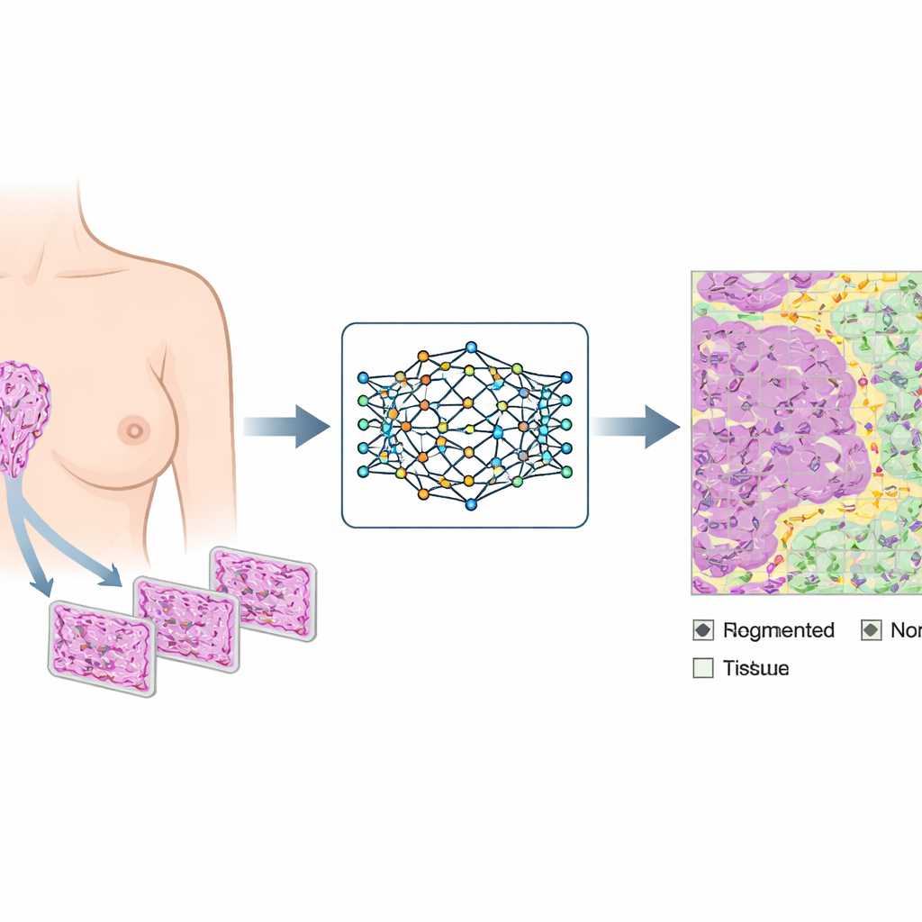
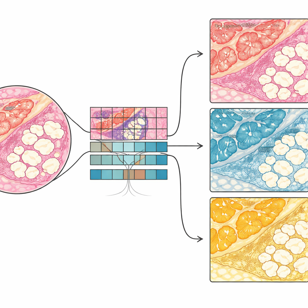
The team assembled 70 high-resolution digital images of normal breast tissue from five institutions in the United Kingdom, the Netherlands, Switzerland and a public tissue bank. These samples came from women aged 16 to 74 with different backgrounds, including healthy volunteers, women having breast reduction surgery, those with inherited high-risk gene changes, and women with cancer in the opposite breast. Expert pathologists carefully marked three key components on each slide: epithelium (the cell layers lining ducts and lobules), stroma (fibrous and connective tissue) and adipocytes (fat cells). This labor-intensive annotation produced a richly varied reference set that reflects real-world differences in tissue processing, staining and scanning.
Teaching the Computer to See Tissue Types
Using this annotated library, the researchers trained deep learning models—called NBT-Classifiers—to recognise the three tissue types by looking at small image patches taken from the larger slides. They tested different patch sizes and technical settings, such as how to standardise stain color and which neural network architecture to use, ultimately finding a combination that worked best. When evaluated on completely separate collections of normal slides from other centers, the models correctly distinguished epithelium, stroma and fat with near-perfect accuracy. Visual “heatmaps” showed that the AI was focusing on biologically meaningful structures such as cell-rich regions, collagen fibers and fat cell borders, mirroring the way human pathologists interpret tissue.
What Makes Normal Tissue Look Normal
To understand whether training only on healthy tissue offered an advantage, the authors compared their model with an existing tool trained on a mix of normal, precancerous and cancerous samples. Both could identify broad tissue types, but the new NBT-Classifiers were better at capturing the fine-grained architecture of truly normal breast epithelium. When challenged with patches that included early lesions and tumors, the normal-only model more reliably separated healthy-looking areas from abnormal ones. This suggests it has learned a sharper definition of normal breast tissue, which may help highlight subtle deviations that accompany early disease.
From Whole Slides to Targeted Regions
Because the models work at the patch level, they can be applied across entire digital slides, automatically coloring each tiny area as epithelium, stroma or fat. The researchers built an end-to-end pipeline that first detects where tissue is present on a slide, then runs the NBT-Classifiers to create detailed maps of tissue compartments. From these maps, the system can locate individual lobules and their immediate surroundings, generate masks for further measurements, and feed selected regions into more advanced analysis tools. This makes it easier to study specific microenvironments—such as the stroma just outside lobules—where early cancer-related changes may appear, and to combine structural features with other data types like spatial gene or protein maps in future work.
What This Means for Future Breast Cancer Prevention
In plain terms, this study shows that AI can be trained to recognise the building blocks of healthy breast tissue with remarkable precision and in a way that pathologists can interpret. By turning huge, complex digital slides into structured maps of epithelial, fibrous and fatty regions, NBT-Classifiers create a reliable baseline for what normal looks like across many women and hospitals. This clearer picture of normality may make it easier to detect the faint footprints of cancer development earlier than is currently possible, supporting future tools that help identify women at higher risk and guide prevention strategies before visible tumors arise.
Citation: Chen, S., Parreno-Centeno, M., Booker, G. et al. Normal breast tissue (NBT)-classifiers: advancing compartment classification in normal breast histology. npj Breast Cancer 12, 41 (2026). https://doi.org/10.1038/s41523-026-00896-2
Keywords: normal breast tissue, computational pathology, deep learning, early cancer detection, digital histology