Clear Sky Science · en
Similar minds age alike: an MRI similarity approach for predicting age-related cognitive decline
Why this research matters for aging brains
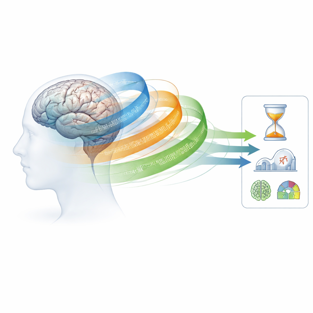
Many people fear losing their memory or thinking skills as they grow older, yet others stay mentally sharp well into their 80s. This study asks a simple but powerful question: can we read the brain’s structure on routine MRI scans to spot who is aging normally and who may be at higher risk of future cognitive problems? The researchers introduce a new way to look at standard brain images that appears to detect the earliest subtle changes of aging—well before traditional brain scans usually show clear damage.
A new way of comparing brain regions
Most brain scans used to study aging focus either on the "wiring" that links regions (anatomical connectivity from diffusion MRI) or on how regions activate together over time (functional connectivity from resting-state fMRI). Both have deepened our understanding of aging, but they are technically demanding, noisy, and not always practical in routine clinics. The authors instead turn to a simpler scan: the standard structural MRI that shows gray matter, the tissue where nerve cell bodies sit. They measure how similar or different brain regions are in their gray matter volume patterns, building what they call gray matter similarity networks. Rather than only asking how thick or large each region is, this method asks how each region statistically resembles every other region, creating a map of structural relationships across the whole brain.
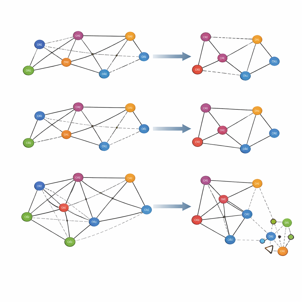
From youthful patterns to individual brain fingerprints
To turn this idea into a practical tool, the team first built a reference network from young adults whose brains represent a "typical" youthful pattern. For each older participant, they then created a slightly altered version of this youthful network by adding that person’s data and measuring how the connections between regions changed. The difference between the reference and the new network becomes a personal deviation map, effectively showing how far each individual’s brain organization has drifted from the young pattern. This approach, tested in more than 800 healthy adults aged 18 to 88 across two independent cohorts, remains computationally efficient while relying on a single, widely available MRI measure: gray matter volume.
Earlier and stronger signals of brain aging
When the researchers asked three different network types—anatomical, functional, and gray matter similarity—to "guess" a person’s age using advanced graph neural networks, the gray matter similarity networks consistently performed best. They predicted age more accurately than either anatomical or functional connectivity and even better than simple measures of gray matter loss. Importantly, markers derived from gray matter similarity started to shift in the early 30s, whereas anatomical networks changed noticeably only in the 40s and functional networks mainly after the late 50s. This suggests that the new method can pick up very early, subtle alterations in how brain regions are structurally related, long before conventional measures detect clear decline. The same pattern held when the authors tested how well each network explained performance on memory, language, movement, emotion, and executive tasks: gray matter similarity features were by far the most informative.
Links to brain cells and thinking abilities
Digging deeper, the team found that the regions most affected in the gray matter similarity networks tended to share particular microscopic tissue features, especially those tied to cortical layers known as II and III. These layers are common in so-called association cortices—areas that integrate information and support complex thinking. They are also thought to be more vulnerable to aging. In contrast, more traditional connectivity measures were most affected in primary sensory areas. The gray matter similarity changes therefore seem to reflect biologically meaningful shifts in the brain’s cellular architecture, not just overall shrinkage. When all three network types were combined into a single multimodal model, predictions improved further, but most of the added power still came from the gray matter similarity component.
What this means for brain health and the future
In everyday terms, this study shows that how similar different parts of your brain look to each other on a routine MRI can reveal how your brain is aging, often years before more obvious damage appears. Gray matter similarity networks provide a kind of early-warning map of brain organization that closely tracks both age and thinking abilities, while remaining robust to many individual differences. Although this work is cross-sectional and needs confirmation in long-term follow-up studies, it points toward a practical, biologically grounded marker that could one day help doctors identify people at risk for age-related cognitive decline or neurodegenerative disease earlier, when prevention and treatment might be most effective.
Citation: Zufiria-Gerbolés, B., Sun, J., Pineda, J. et al. Similar minds age alike: an MRI similarity approach for predicting age-related cognitive decline. npj Aging 12, 39 (2026). https://doi.org/10.1038/s41514-026-00345-1
Keywords: brain aging, MRI, cognitive decline, brain networks, neuroimaging biomarkers