Clear Sky Science · en
Parkin deficiency impairs female fertility, oocyte development, fertilization and mitochondrial function in mice
Why the Health of Egg Cells Matters
Many people today are choosing to have children later in life, but female fertility naturally declines with age. One major reason is that egg cells, which must power early embryo development, depend heavily on tiny energy factories called mitochondria. This study explores a little-known protector of mitochondrial health, a protein called Parkin, and asks a simple question: what happens to female fertility when Parkin is missing?
A Cell Cleanup Crew with a New Job
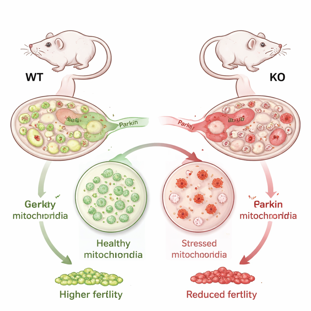
Parkin is best known for its role in brain cells, where it helps remove damaged mitochondria and is linked to certain forms of Parkinson’s disease. Until now, its role in the ovary and egg cells had been largely overlooked. The researchers suspected that because egg cells need so much energy, they might be especially vulnerable if this cleanup system fails. Using mice that completely lack Parkin, they examined how well females could produce eggs, how often those eggs were fertilized, and what was happening inside the cells at the molecular level.
Fewer Eggs and Lower Chance of Fertilization
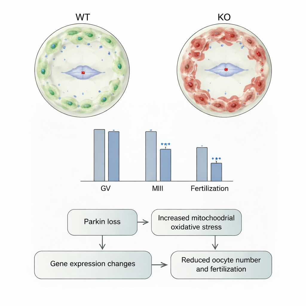
When Parkin-deficient female mice were housed with healthy males for three months, they had noticeably smaller families: on average, each produced about one-third fewer pups than normal females. Looking more closely, the team stimulated the ovaries to release egg cells and counted them at two key stages. In young adult mice, those without Parkin produced far fewer immature eggs and far fewer mature eggs ready for fertilization compared with ordinary mice. Similar, though slightly weaker, trends appeared in older animals. When the researchers performed in vitro fertilization, eggs from Parkin-deficient females were significantly less likely to become fertilized, showing that the problem was not just egg number but egg quality.
Not Fewer Follicles, but Poorer-Performing Eggs
The ovaries store eggs inside tiny structures called follicles, which progress from resting to fully grown over time. The scientists examined thin sections of ovaries under the microscope and counted follicles at all stages, from the earliest primordial follicles to fully developed antral follicles. Surprisingly, they found no meaningful difference between mice with and without Parkin: the basic ovarian “stock” of follicles appeared intact. This suggests that Parkin is not required to build or maintain the reserve of eggs, but is instead crucial later on, when eggs grow, mature, and prepare for fertilization.
Stressed Mitochondria and Shifting Gene Activity
To understand what was going wrong inside the eggs, the team measured mitochondrial stress and gene activity. In immature eggs from Parkin-deficient mice, mitochondria produced more reactive oxygen species—chemically reactive byproducts that can damage cell components—indicating higher oxidative stress. However, the overall electrical charge across the mitochondrial membrane, a sign of basic energy production, was not noticeably changed. The researchers also extracted and sequenced RNA from eggs at several ages to see which genes were turned up or down. Dozens of genes showed altered activity in Parkin-deficient eggs, and the number grew with age. Many of these genes were involved in protein production, stress responses, and energy metabolism, suggesting that ongoing mitochondrial stress slowly reshapes how the egg cell functions.
What This Means for Fertility and Aging
Taken together, the findings show that Parkin is a key guardian of egg cell health in mice. Without it, eggs experience more mitochondrial stress, their internal machinery changes, fewer eggs reach maturity, and fewer can be successfully fertilized, even though the overall reserve of follicles in the ovary looks normal. For a layperson, this means that fertility is not only about how many eggs a female has, but also about how well those eggs’ energy systems are maintained over time. While this work was done in mice, it raises the possibility that similar mitochondrial quality-control pathways, including Parkin, may influence age-related infertility in women and could one day become targets for new treatments or diagnostic tools.
Citation: Volovsky, M., Rodríguez-Eguren, A., Ergun, Y. et al. Parkin deficiency impairs female fertility, oocyte development, fertilization and mitochondrial function in mice. npj Aging 12, 33 (2026). https://doi.org/10.1038/s41514-026-00332-6
Keywords: female fertility, mitochondria, Parkin protein, oocyte quality, reproductive aging