Clear Sky Science · en
Identification of altered immune landscape at single-cell resolution in NSCLC brain metastasis and its association with poor immune checkpoint inhibitor responses
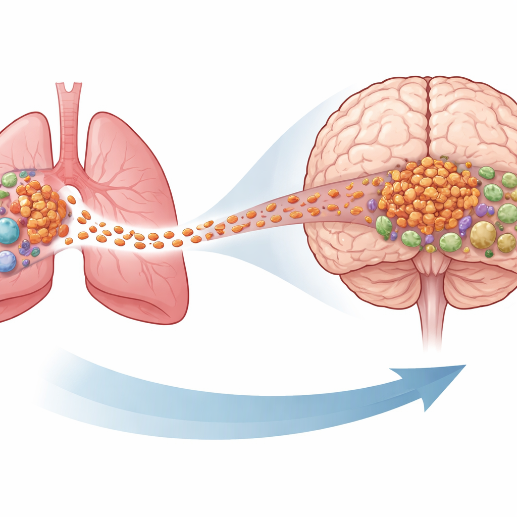
Cancer’s Spread to the Brain and Why Treatment Often Fails
When lung cancer spreads to the brain, doctors can now use powerful drugs that unleash the immune system, called immune checkpoint inhibitors. Yet many patients with these medicines still see their brain tumors grow or return, even when tumors in the lung respond. This study asks a simple but crucial question: what is different about the immune cells inside brain metastases compared with those in the original lung tumor, and how might those differences explain why modern immunotherapy so often falls short in the brain?
Looking at Each Immune Cell One by One
To tackle this problem, the researchers used single-cell RNA sequencing, a technology that reads out which genes are active in tens of thousands of individual cells. They collected immune cells from lung tumors and from brain metastases in people with non-small cell lung cancer, the most common form of lung cancer. By profiling more than one hundred thousand cells, they built a high-resolution map of the immune landscape in each location. This allowed them to identify distinct families of T cells, dendritic cells, monocytes, macrophages, B cells, and plasma cells, and to see which cell types were abundant or scarce in the brain compared with the lung.
Stressed T Cells and the Loss of Immune Memory
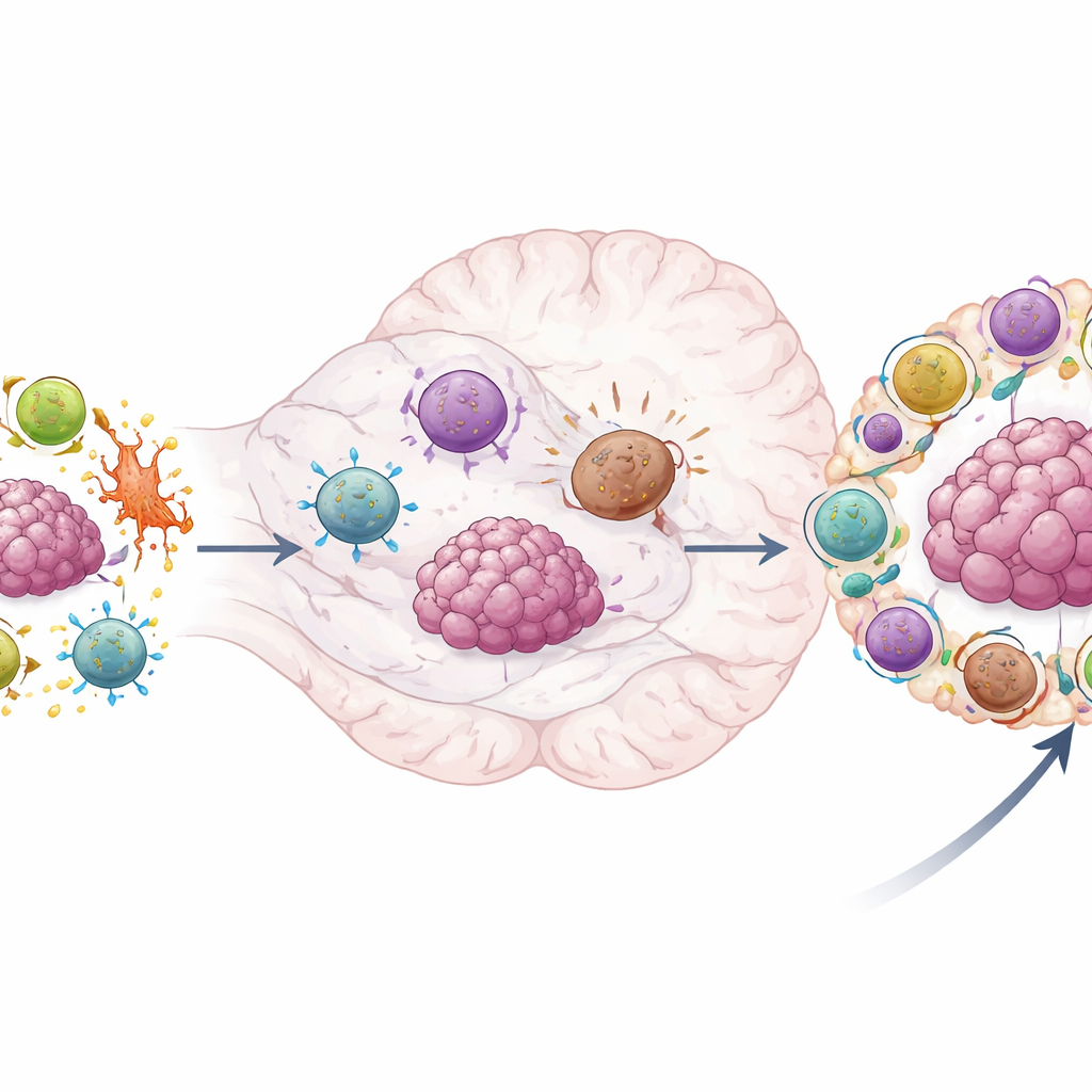
The team found that brain metastases are packed with T cells showing signs of intense cellular stress. These cells switched on high levels of genes that produce a heat‑shock protein called HSP70, a classic marker of cells under pressure. Both helper T cells and killer T cells with this stressed profile were more common in brain lesions than in primary lung tumors. Although some of these cells still carried features of activation or attack, they also bore hallmarks of exhaustion and dysfunction. When the authors analyzed large clinical datasets of patients treated with checkpoint inhibitors, people whose tumors had higher signatures of these HSP70‑high T cells tended to experience faster disease progression.
Equally important, several T cell types that normally act as the long‑lasting “memory” arm of the immune system were depleted in brain metastases. Central memory–like helper T cells and tissue‑resident memory killer T cells, both capable of persisting and rapidly responding to cancer, were more common in lung tumors and were linked to better outcomes after immunotherapy. In the brain, these protective memory pools were replaced by cycling, highly proliferative T cells that showed metabolic strain and were associated with worse responses to treatment. Together, this paints a picture of the brain metastasis as an environment where durable, high‑quality immune surveillance is lost and replaced by stressed, overworked, and less effective T cells.
Support Cells That Help or Hinder the Immune Attack
The story does not stop with T cells. The authors also examined other immune cell types that shape the tumor environment. In primary lung tumors, they found plentiful dendritic cells of a subtype specialized in presenting foreign material to T cells, effectively acting as teachers that train T cells to recognize and fight cancer. These cells were much rarer in brain metastases, and their presence in lung tumors correlated with better survival on checkpoint therapy. Monocytes and macrophages also split into helpful and harmful factions. One monocyte group in lung tumors showed signs of active inflammation and support for immune attack, whereas a different monocyte subset, enriched in the brain, displayed altered energy use suggestive of an immunosuppressive role. Likewise, a macrophage subtype marked by a molecule called PLTP accumulated in brain metastases and was tied to poorer outcomes, while another macrophage group in lung tumors was linked to more favorable responses.
Building a Gene Signature to Predict Treatment Response
By combining their single‑cell discoveries with bulk gene‑expression data from larger patient cohorts, the researchers distilled a seven‑gene “brain metastasis–derived immune signature,” or BMIS. This signature captures the balance between stressed T cells and suppressive macrophages on one side and more effective immune programs on the other. When they applied BMIS to independent groups of patients with lung cancer and metastatic bladder cancer receiving checkpoint inhibitors, higher BMIS scores consistently marked those with worse survival and lower response rates. Notably, the BMIS added predictive power beyond the commonly used tumor mutational burden, suggesting that understanding the state of the immune microenvironment can complement DNA‑based biomarkers.
What This Means for Patients and Future Therapies
For a lay reader, the takeaway is that brain metastases from lung cancer are not just ordinary tumors in a new location; they sit in a profoundly altered immune neighborhood. Key defenders—memory T cells and antigen‑presenting dendritic cells—are depleted, while stressed T cells, metabolically rewired monocytes, and suppressive macrophages dominate. This skewed immune ecosystem helps explain why modern checkpoint drugs often control disease in the lung but falter in the brain. By pinpointing the specific cell types and gene programs involved, and by translating them into a practical gene‑based score, this work lays groundwork for better predicting who will benefit from immunotherapy and for designing new treatments aimed at restoring a healthy, effective immune presence in the brain.
Citation: Bai, M., Yin, T., Li, X. et al. Identification of altered immune landscape at single-cell resolution in NSCLC brain metastasis and its association with poor immune checkpoint inhibitor responses. Nat Commun 17, 2370 (2026). https://doi.org/10.1038/s41467-026-70715-6
Keywords: non-small cell lung cancer, brain metastasis, tumor immune microenvironment, immunotherapy resistance, single-cell RNA sequencing