Clear Sky Science · en
Fragmentomic liquid biopsy enables early breast cancer detection, molecular subtyping and lymph node assessment
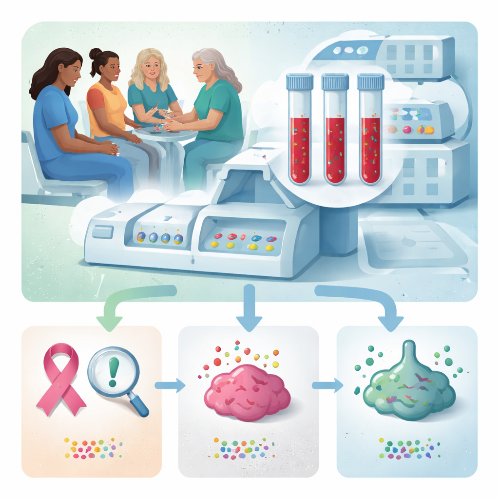
Why a Blood Test for Breast Cancer Matters
Breast cancer is common, but today’s screening tools—like mammograms and ultrasounds—can miss early tumors, especially in women with dense breast tissue. This study explores a different approach: reading tiny fragments of DNA that circulate in the blood to detect cancer, classify its type, and estimate whether it has spread to nearby lymph nodes. If such a test can be made reliable and affordable, it could complement imaging and bring high‑quality screening to more women, including those living far from major hospitals.
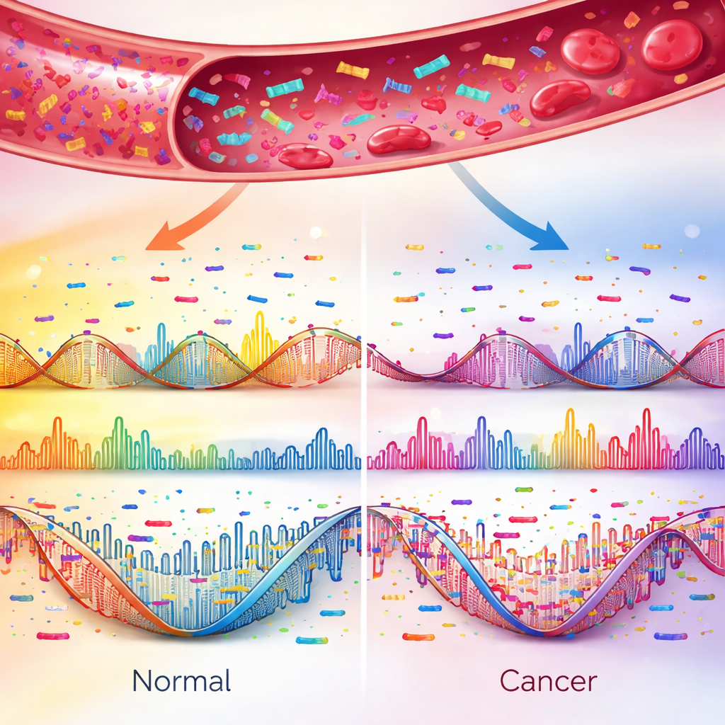
Looking at DNA Dust in the Blood
When cells die, they shed broken pieces of DNA into the bloodstream. Most come from healthy cells, but cancers release their own fragments with telltale patterns. The researchers developed a method called TuFEst that doesn’t search for specific gene mutations. Instead, it examines the “fragmentome”: the sizes of DNA pieces, the short sequence patterns at their ends, and where they fall across the genome. Because these patterns reflect how DNA is packaged and controlled inside cells, cancer cells leave a fragmentation fingerprint that can be picked up with low‑depth whole‑genome sequencing of a small blood sample.
A Large Real‑World Test in Hospitals
The team ran a multicenter study in China, enrolling 503 women with breast cancer—most at very early stages—and 289 women with benign breast conditions. From about one milliliter of plasma per person, they sequenced cell‑free DNA at ultra‑low coverage and fed dozens of fragment features into several machine‑learning models. A stacked ensemble model, which blends the strengths of multiple algorithms, emerged as the best performer and was named TuFEst. It correctly identified 95 percent of cancers while wrongly flagging about 22 percent of non‑cancer cases in the main dataset, and its performance remained strong in independent hospital cohorts.
Finding Hidden Cancers and Tumor Types
To test whether the blood signal could catch cancers that imaging missed, the researchers examined 26 women whose breast lesions had been labeled “probably benign” on both ultrasound and mammography, but who were later found to have invasive cancer after the lesion grew. Using the blood drawn at the time of the original scans, TuFEst correctly called 25 of these 26 cancers. The team then expanded the framework to two related tools. One, TuFEst‑MS, used the same fragmentomic information to sort tumors into common molecular subtypes, such as hormone‑receptor‑positive, HER2‑positive, and triple‑negative disease. It reached around 90 percent accuracy in both training and validation groups, and it matched the subtype of metastatic lesions in most advanced patients, including cases where the metastasis differed from the original tumor.
Clues About Cancer Spread and Behavior
A third model, TuFEst‑LN, aimed to flag whether cancer had spread to lymph nodes in the armpit—an important factor in choosing surgery and drug treatment. In women whose node status was known from surgery, the blood‑based tool distinguished node‑positive from node‑negative cases with good accuracy and, crucially, a very high negative predictive value: over 90 percent in the main validation group and 97.6 percent in especially tricky cases where imaging and pathology disagreed. High “cancer scores” from TuFEst also aligned with more aggressive tumor biology. By analyzing RNA from 79 matched tumor samples, the authors showed that high‑score cancers were enriched for fast growth, inflammatory signaling, and immune‑active microenvironments, patterns often seen in HER2‑positive and triple‑negative breast cancers.
What This Could Mean for Patients
For non‑specialists, the takeaway is that a simple blood draw may one day help do three things at once: detect breast cancer early, indicate its biological subtype, and suggest whether it has reached the lymph nodes—all without additional imaging or invasive biopsies in many cases. The test still needs prospective trials in broader screening settings, and it is not yet a replacement for mammograms or ultrasounds. But this work shows that the “dust” of DNA fragments in our blood carries surprisingly rich information, and that smart analysis of these patterns could support more timely, less invasive, and more personalized breast‑cancer care.
Citation: Zhu, Y., Zheng, S., Shao, Y. et al. Fragmentomic liquid biopsy enables early breast cancer detection, molecular subtyping and lymph node assessment. Nat Commun 17, 2276 (2026). https://doi.org/10.1038/s41467-026-70204-w
Keywords: breast cancer, liquid biopsy, cell-free DNA, early detection, machine learning