Clear Sky Science · en
Targeting tRNA-dependent tyrosine usage unveils a metabolic vulnerability in hepatocellular carcinoma
Starving Cancer Cells of a Common Building Block
Liver cancer cells, like all rapidly dividing cells, need a steady supply of amino acids—the tiny building blocks of proteins. This study reveals that hepatocellular carcinoma, the most common form of liver cancer, has an unexpected weakness: it depends heavily on the amino acid tyrosine for both energy production and survival. By selectively limiting how cancer cells use tyrosine, the researchers were able to slow tumour growth, damage cancer cell power plants, and trigger a form of cell death that may be harnessed for new treatments.
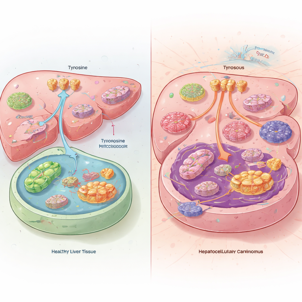
A Hidden Imbalance in Liver Tumours
When the team compared liver tumours with healthy liver tissue from patients and mice, they found something puzzling. Inside the cancer cells, tyrosine levels were actually low, even though the tumours were pulling in more tyrosine from the bloodstream and breaking down less of it. Instead of burning tyrosine as fuel, liver tumours re-routed it into protein production. The cancers achieved this by increasing the activity of tyrosine transporters at the cell surface and turning down the enzymes that normally dismantle tyrosine in the liver. Mice fed a tyrosine-free diet developed smaller tumours and lived longer, while extra tyrosine in the diet made their cancers grow faster.
How Cancer Rewires Protein Production
The researchers traced this tyrosine hunger to a powerful cancer-driving gene called MYC. In liver tumours, MYC switches on a protein called YARS1, which attaches tyrosine to a specific transfer RNA (tRNA-TyrGUA). This loaded tRNA is what ribosomes need to build tyrosine-rich proteins. The study showed that liver cancers boost both YARS1 and its matching tRNA, ensuring a privileged pipeline of tyrosine into protein synthesis. Disrupting this pipeline—either by lowering tyrosine levels, knocking down YARS1 or tRNA-TyrGUA, or blocking tyrosine loading—made liver cancer cells far less fit in dishes and in mice, revealing a metabolic vulnerability tightly linked to their high protein production needs.
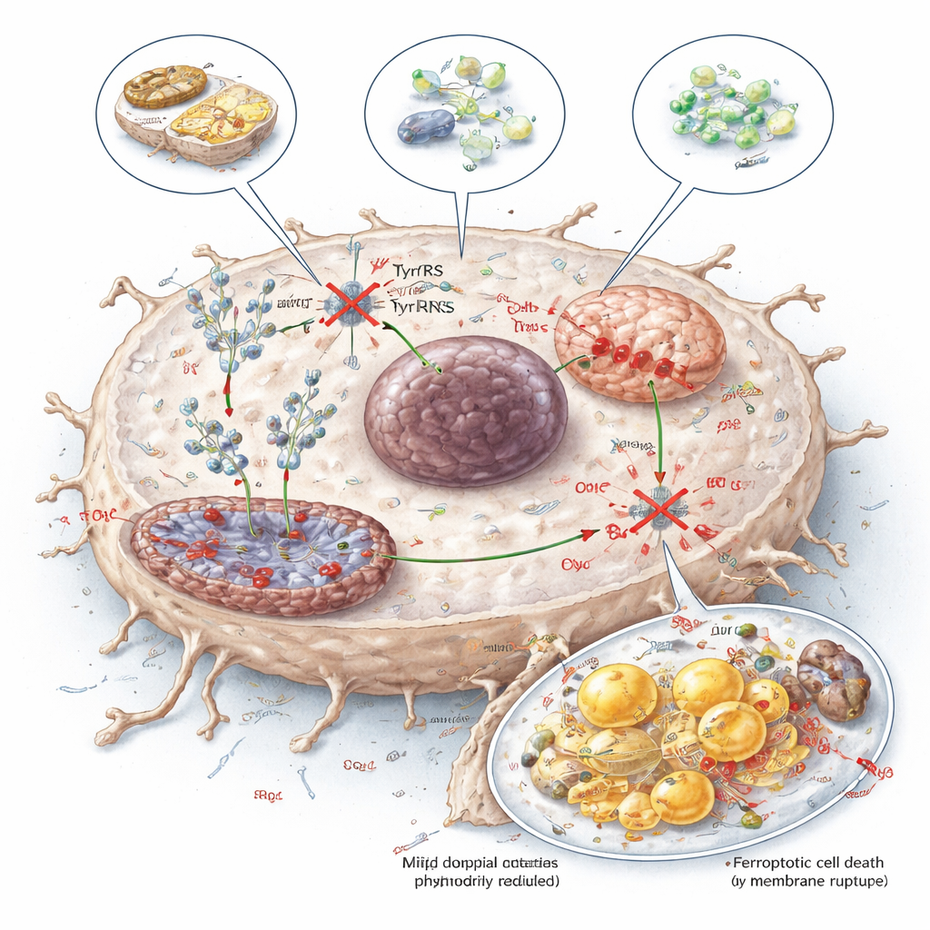
Crippling the Powerhouses and Lipid Balance
By combining genome-wide measurements of RNA, protein production, and metabolites, the team identified two especially sensitive targets of tyrosine restriction. One, NDUFB8, is a component of mitochondrial complex I, a core part of the cell’s energy-generating machinery. The other, SCD1, helps convert saturated fats into monounsaturated fats that are safer to store in cell membranes and droplets. When tyrosine for translation was restricted, the production of NDUFB8 and SCD1 dropped, while their RNA levels stayed largely unchanged—clear evidence that translation, not gene expression, was being hit. This led to poorly assembled complex I, reduced cellular respiration, excess reactive oxygen species, and a shift in fats from monounsaturated toward more fragile polyunsaturated forms that are easily oxidized.
Forcing Cancer Cells into an Iron-Driven Death
The combined mitochondrial damage and unstable fats pushed liver cancer cells toward ferroptosis, an iron-dependent form of cell death driven by lipid peroxidation. The tumours tried to fight back by increasing natural ferroptosis blockers such as GPX4 and related molecules, but a large CRISPR gene-editing screen showed that disabling these protectors made tyrosine restriction even more lethal. In cell and mouse models, pairing tyrosine-limiting strategies with drugs that inhibit GPX4 or BCL2—or with existing liver cancer drugs like sorafenib and venetoclax—produced stronger tumour control, smaller tumour burdens, and longer survival.
Turning a Metabolic Weakness into Therapy
To explore practical ways of exploiting this weakness, the researchers tested three approaches: a tyrosine-restricted diet, an enzyme (TAL) that enzymatically degrades tyrosine, and a small molecule called tyrosinol that competes with tyrosine for binding to YARS1. All three reduced the availability of tyrosine for protein synthesis, lowered NDUFB8 and SCD1 levels, damaged mitochondrial function, and increased ferroptosis in liver tumours, while showing manageable effects on normal tissues in mice. For a layperson, the message is that liver cancers appear to be hooked on tyrosine not just as a nutrient, but as a precise fuel for their protein-making machinery. Targeting this dependency—by diet, enzymes, or drugs—offers a promising new way to weaken tumours and boost the impact of existing treatments.
Citation: Zhang, H., Wang, Z., Zhao, Y. et al. Targeting tRNA-dependent tyrosine usage unveils a metabolic vulnerability in hepatocellular carcinoma. Nat Commun 17, 2244 (2026). https://doi.org/10.1038/s41467-026-70112-z
Keywords: liver cancer, amino acid metabolism, tyrosine, mitochondria, ferroptosis