Clear Sky Science · en
Transcranial focused ultrasound induces source localizable cortical activation in resting state humans when applied concurrently with transcranial electric stimulation
Fine-Tuning the Brain Without Surgery
Imagine being able to gently nudge a very precise spot in the brain without cutting into the skull—helping doctors treat depression, epilepsy, or movement problems with fewer side effects. This study explores whether two noninvasive methods—weak electrical currents on the scalp and carefully aimed ultrasound waves—can be combined to selectively "wake up" a small patch of human cortex while a person is simply resting with eyes closed. The work helps resolve a major debate: does focused ultrasound truly act on specific brain areas, or are its effects mostly a side effect of sound in the ears?
Two Different Ways to Nudge Nerve Cells
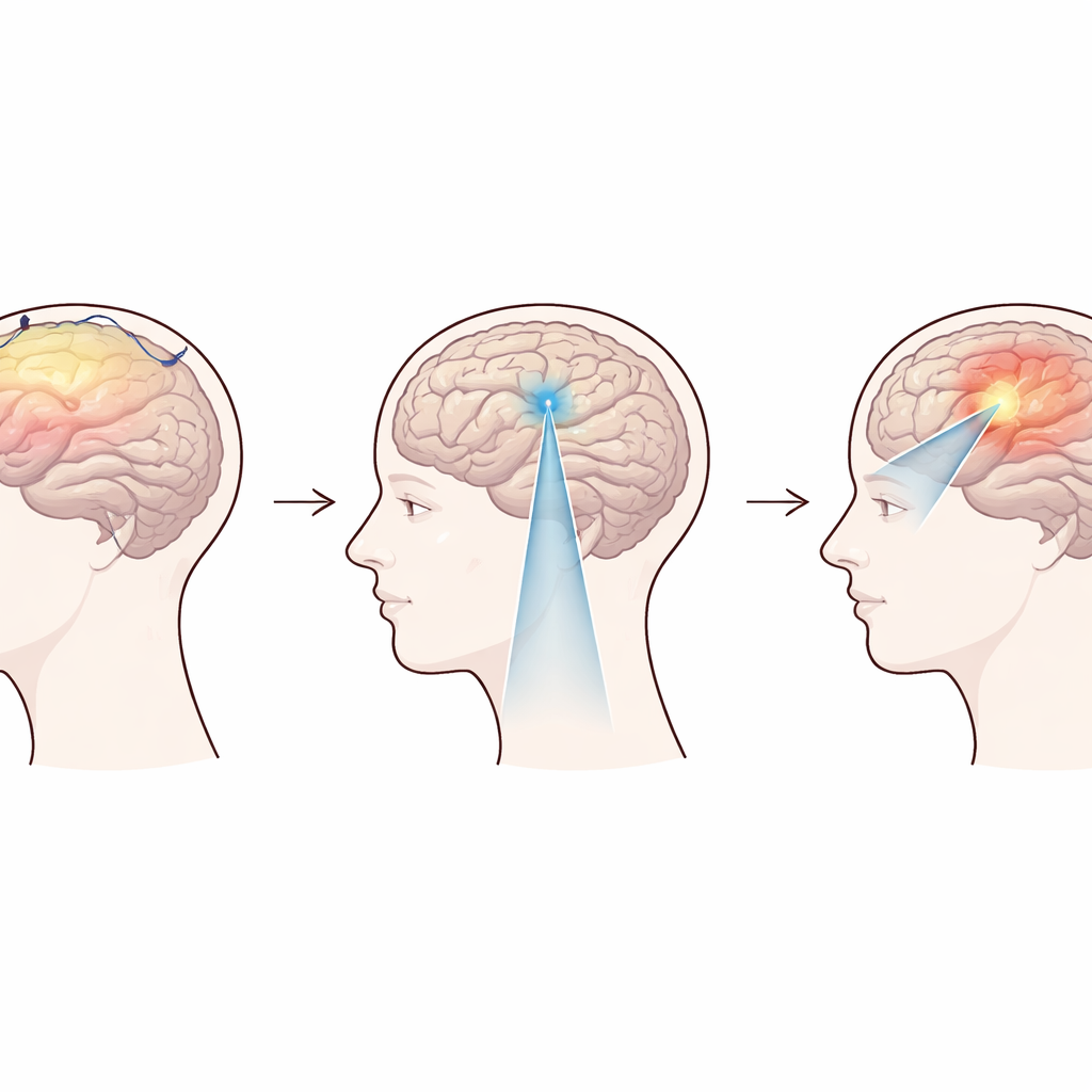
The researchers worked with two tools that each influence the brain in a different way. Transcranial direct current stimulation (tDCS) passes a very weak, steady electrical current between scalp electrodes. On its own, it usually does not trigger brain cells to fire; instead it makes them slightly more or less likely to respond to other inputs. Transcranial focused ultrasound (tFUS), by contrast, sends sound waves through the skull that can be focused on small regions only a few millimeters wide. Animal and human work has suggested that these pressure waves may tug on tiny mechanical sensors in cell membranes, subtly changing how easily neurons activate. The central question was whether tFUS by itself can reliably trigger activity in a chosen patch of human cortex, or whether its main effect is simply to create an audible pulsing that activates the hearing system.
A New Blend: Electricity Plus Ultrasound
The team studied 27 healthy volunteers at rest, recording their brain activity with whole-head electroencephalography (EEG). They tested three main conditions targeted at the left motor cortex, the region that helps control movements of the right hand. In one, they applied tDCS alone. In another, they applied tFUS alone, with different pulse patterns meant to either excite or inhibit neurons. In the third, called transcranial electro-acoustic stimulation (tEAS), they applied tDCS and tFUS at the same time, so that a weak electric shift and a mechanical push would reach the same group of neurons together. They also used control setups that aimed ultrasound at a different brain region to separate true local effects from whole-brain or auditory responses.
What the Brain Signals Revealed
EEG allowed the scientists to look not just at surface signals, but to mathematically reconstruct where inside the brain those signals likely came from. When they examined the first 200 milliseconds after each stimulus, they found that tFUS alone produced clear, repeatable peaks in activity at about 30, 90, and 170 milliseconds. However, these responses were spread symmetrically across both sides of the head and traced back mainly to the hearing areas and deeper structures—not to the targeted motor cortex. Strikingly, the same kind of response appeared whether the ultrasound was aimed at motor cortex or at prefrontal cortex, and advanced statistical tests supported that these patterns were essentially the same. Additional connectivity analysis showed that ultrasound increased information flow from primary auditory cortex to deep brain regions, even when the pulse rate was too high to be consciously heard. In short, the strong EEG signals from tFUS alone looked and behaved like auditory-driven responses, not focal cortical stimulation.
When Two Gentle Pushes Add Up
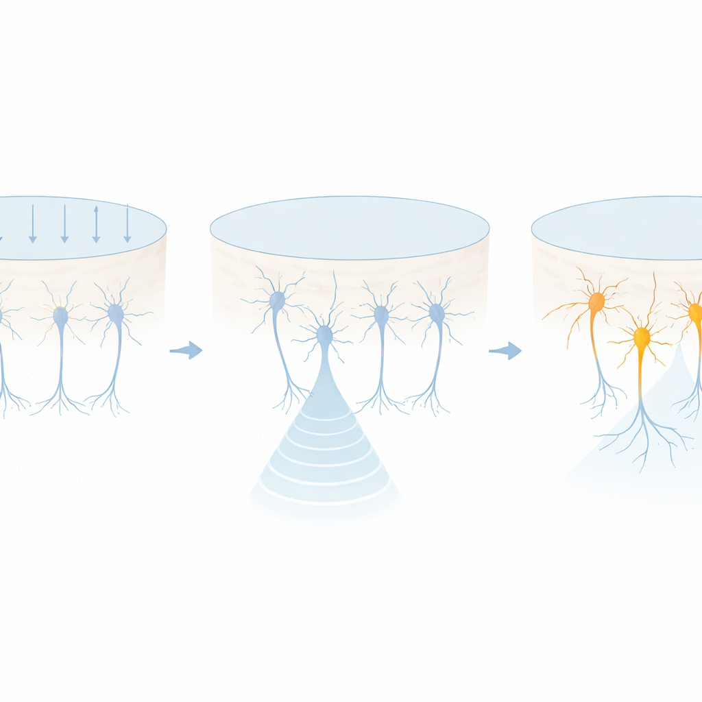
The picture changed when tDCS and tFUS were combined as tEAS. In these trials, EEG source imaging showed a robust and statistically significant boost of activity in the motor cortex region directly under the stimulation site, compared with the same patch on the opposite side of the brain. This focused response appeared for both excitatory and inhibitory tDCS polarities, with the sign of the EEG signal flipping depending on how the current was oriented. Importantly, neither tDCS alone nor tFUS alone produced such localized, source-resolvable activation under the same resting conditions, and simply adding together their separate effects in the analysis could not reproduce the tEAS pattern. The authors also broadened the ultrasound settings—changing pulse rates, duty cycles, and even increasing in-brain pressure within safety limits—and still found no evidence that tFUS by itself drove a clear, site-specific cortical response in resting humans.
A New View of How Ultrasound Shapes the Brain
To make sense of these findings, the researchers turned to a classic mathematical model of nerve cells, the Hodgkin–Huxley model, and added a pathway representing mechanically sensitive ion channels. Simulations showed that a subthreshold mechanical effect (from ultrasound) and a subthreshold electrical shift (from tDCS) can combine to cross the firing threshold and generate full action potentials. This matches the experimental observation that only the combined tEAS condition produced focal, source-localizable cortical activation. The authors argue that in humans, at safe pressure levels, focused ultrasound most likely acts as a subthreshold co-modulator: it changes the readiness of neurons to respond, but typically needs another input—such as tDCS, sensory stimulation, or attention—to drive strong, location-specific firing. This framework helps reconcile why some studies see powerful behavioral effects of tFUS when it is paired with other tasks or stimuli, while others see mainly auditory-related activity when it is used alone.
Citation: Kosnoff, J., Gonsisko, C., Yu, K. et al. Transcranial focused ultrasound induces source localizable cortical activation in resting state humans when applied concurrently with transcranial electric stimulation. Nat Commun 17, 2023 (2026). https://doi.org/10.1038/s41467-026-69853-8
Keywords: transcranial focused ultrasound, noninvasive brain stimulation, EEG source imaging, neuromodulation, tDCS