Clear Sky Science · en
Phenotype of circulating tumor-reactive T cells predicts immune checkpoint inhibitor response in non-small cell lung cancer
Why blood-based cancer clues matter
Most people know that the immune system can sometimes recognize and attack cancer. What is far less obvious is that important clues about whether a patient will benefit from modern immunotherapy may be hiding in a simple blood draw. This study focuses on a rare group of immune cells in the bloodstream of people with non-small cell lung cancer and shows that their "look" and behavior can forecast how well immune checkpoint drugs will work.
Special hunter cells in the blood
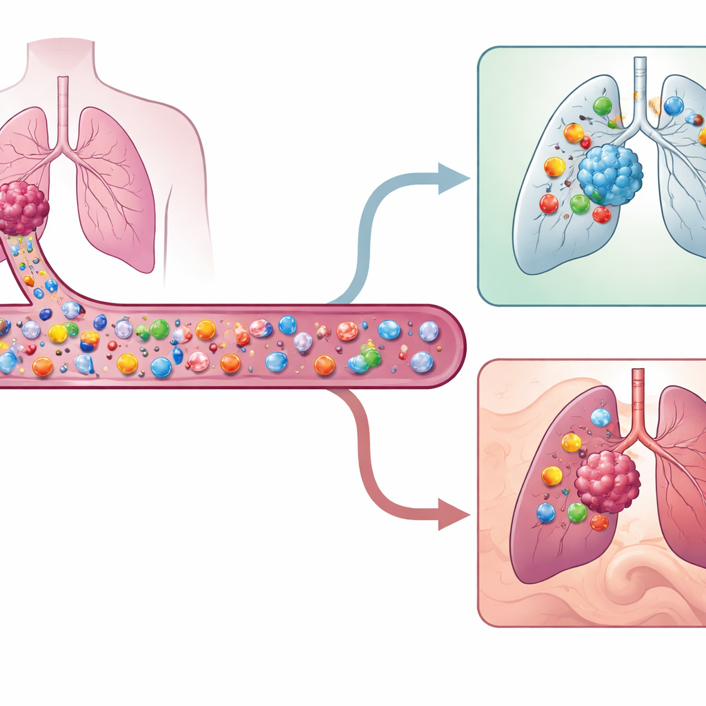
Cancers carry molecular flags that mark them as abnormal. Certain white blood cells, called T cells, can recognize these flags and kill cancer cells. But not all T cells inside a tumor actually recognize the cancer; many are just bystanders reacting to past infections. The researchers set out to find and characterize the true cancer-hunting T cells that are circulating in the blood rather than already sitting in the tumor. These circulating tumor-reactive T cells are extremely rare—often less than one in a thousand T cells—so the team used high-resolution single-cell genetic and protein profiling to spot them in nine patients with early-stage non-small cell lung cancer.
A fingerprint for cancer-fighting T cells
To link blood T cells to those inside tumors, the scientists used each T cell’s unique receptor sequence as a kind of barcode. If a blood T cell shared the same receptor as a tumor-infiltrating cell with a known tumor-reactive gene pattern, it was tagged as tumor-reactive in the blood. These circulating cells showed a distinctive surface fingerprint: they tended to carry proteins called CD49a, CD49b and HLA-DR, and to lack a protein associated with naïve cells, CD45RA. Together, this combination—high CD49a or CD49b, high HLA-DR, and low CD45RA—marked a previously unrecognized subset of activated, tissue-seeking memory T cells that are primed to enter tumors. The team also derived a 140-gene signature that separates these rare hunters from other blood T cells more accurately than earlier methods developed for other cancers.
From early-stage scouts to exhausted fighters
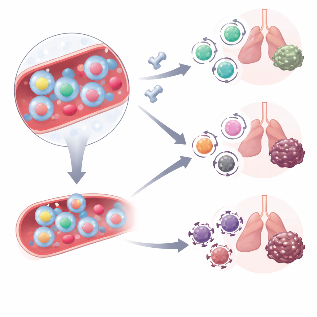
By reconstructing likely developmental paths from blood to tumor, the researchers found that circulating tumor-reactive T cells look like progenitors—earlier-stage cells—with higher levels of genes linked to long-term survival and lower levels of exhaustion-related genes than their counterparts already inside tumors. Once in the tumor, these cells gradually acquire more signs of tiredness and tissue residency. Interestingly, even in the blood they already show features of cells adapted to live in tissues, suggesting that they are poised to home in on inflamed or cancerous sites rather than wander broadly through the body.
How these cells change with treatment
The team next asked how these blood-borne cancer hunters behave when patients receive immune checkpoint inhibitors that block the PD-1 pathway. They analyzed blood T cells from another group of lung cancer patients before and just after starting combination PD-1 blockade and chemotherapy. Before treatment, both responders and non-responders had similar numbers of circulating tumor-reactive cells, but their quality differed. In non-responders, these cells more often carried high levels of CD38, a molecule linked to metabolic stress and resistance to PD-1 therapy. After the first treatment dose, tumor-reactive cells in responders shifted toward a stem-like effector memory state—cells that combine killing ability with the capacity to persist—while in non-responders they remained in a more highly activated state.
Animal tests and real-world validation
To confirm that these markers truly flag cancer-specific cells, the scientists used a mouse melanoma model engineered to express an artificial target that can be tracked with a molecular "tetramer" probe. In these mice, T cells that recognized the artificial target in blood were strongly enriched among cells bearing the same surface marker trio seen in human patients. After PD-L1 blockade, these mouse tumor-reactive cells lost some of their activation markers while remaining functionally engaged, mirroring the shift seen in human responders. Finally, in a larger group of 70 lung cancer patients receiving checkpoint therapy, two blood measurements stood out: patients who started treatment with fewer CD38-high tumor-reactive cells and who showed a drop in the activation-marker–defined population after the first dose enjoyed much longer periods before their cancer progressed.
What this means for patients
In everyday terms, this work shows that a tiny set of "elite" cancer-hunting T cells in the bloodstream carries important hints about how a person will respond to immunotherapy. It is not how many of these cells are present that matters most, but whether they are metabolically healthy and able to shift into a long-lived, stem-like state once checkpoint drugs release their brakes. Simple blood tests that look at the surface patterns and activation state of these rare cells could, with further refinement, help doctors predict who is most likely to benefit from immune checkpoint inhibitors and guide the development of new T cell–based treatments drawn directly from the blood.
Citation: Ito, K., Iida, K., Hirano, T. et al. Phenotype of circulating tumor-reactive T cells predicts immune checkpoint inhibitor response in non-small cell lung cancer. Nat Commun 17, 2856 (2026). https://doi.org/10.1038/s41467-026-69680-x
Keywords: tumor-reactive T cells, immune checkpoint therapy, non-small cell lung cancer, blood biomarkers, T cell exhaustion