Clear Sky Science · en
Pyrimidinergic calcium signaling links tubular metabolism to fibrosis in kidney disease
Why kidney scarring matters
Chronic kidney disease quietly affects hundreds of millions of people worldwide and often ends with the kidneys becoming scarred and stiff. Once this scarring, called fibrosis, takes hold, lost kidney function is hard to regain. Yet doctors still lack drugs that specifically stop fibrosis. This study asks a basic but crucial question: how does short-term damage to the kidney’s filtering tubes gradually turn into lasting scar tissue, and could interrupting that chain of events slow or prevent kidney failure?
Busy kidney tubes and their hidden chemistry
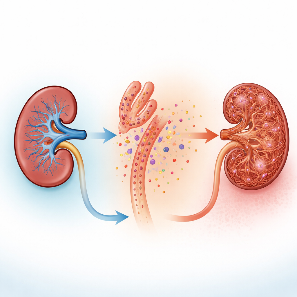
The kidney’s proximal tubules are microscopic workhorses that reclaim most of the water and nutrients filtered from blood. To power this job, their chemistry is unusually active. By analyzing single-cell gene expression data from mouse kidneys, the researchers found that these tubule cells are especially rich in genes involved in handling pyrimidines, a family of small molecules that normally help build and repair DNA and RNA. In injured tubules, one enzyme in a “salvage” pathway, cytidine deaminase, was turned up sharply, suggesting that damaged cells reorganize pyrimidine use to keep certain energy-rich building blocks, including a compound called UDP, topped up.
Signals that spill out of damaged cells
When the team injured human tubule-like cells in culture with toxins that mimic chemotherapy or folic acid overdose, the cells released UDP into the fluid around them. In actual mouse kidneys exposed to injury, staining of tissue slices showed both high levels of the pyrimidine-handling enzyme in tubules and signs that neighboring support cells, called fibroblasts, were waking up and changing shape. Fibroblasts normally sit quietly between tubules, but when activated they multiply and help lay down collagen and other fibers that thicken and stiffen kidney tissue. These observations suggested a simple idea: injured tubules might be “spilling” chemical distress signals that neighboring fibroblasts can sense.
How fibroblasts listen with calcium flashes
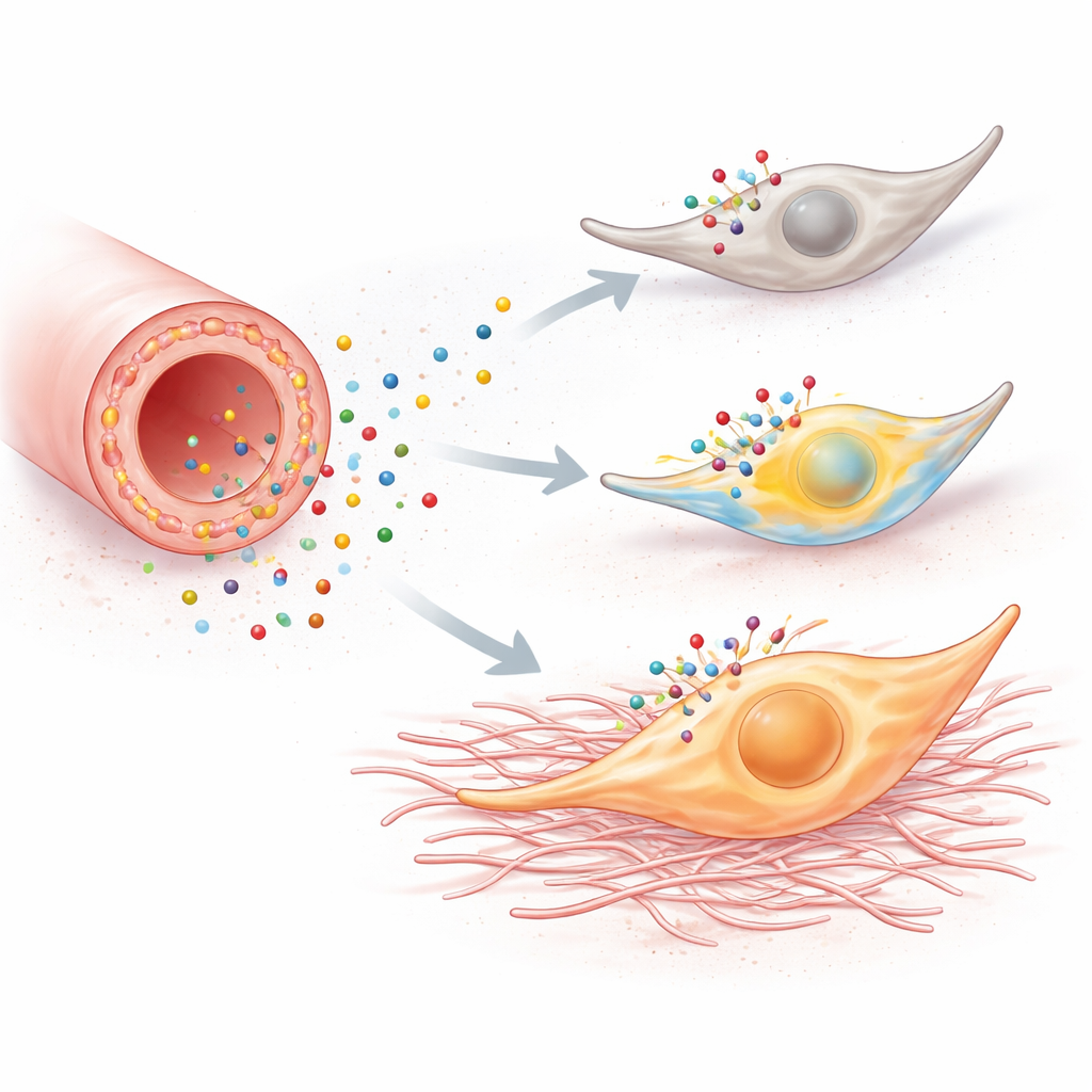
The suspected “ears” on fibroblasts are P2Y6 receptors, surface proteins that respond to UDP. Mining several single-cell datasets, the authors saw that P2Y6 appeared on stromal cells and rose further in mouse models of chronic kidney disease. Measurements across whole kidneys confirmed strong increases in P2Y6 levels in two different scarring models. In fresh kidney slices and in cultured kidney fibroblasts engineered to light up when calcium inside the cell rises, adding UDP or a P2Y6-activating drug produced fast calcium surges. These flashes started in the long, thin processes that wrap around tubules and then swept into the cell body, and they disappeared when P2Y6 was blocked or calcium was chemically soaked up. In live mice, high-resolution intravital microscopy showed that fibroblasts around tubules display frequent, irregular calcium activity that ramps up dramatically when tubules are injured, even as the tubule cells themselves fall silent.
From brief signals to lasting scars
Calcium bursts inside a cell are not just fireworks; they can reprogram behavior. When fibroblasts in culture were bathed in a P2Y6-activating compound, they multiplied faster, crawled more readily, and switched on genes associated with a more aggressive, “myofibroblast” state. These genes encode proteins such as fibronectin, vimentin, and collagen that directly contribute to scar formation. Blocking P2Y6 or preventing calcium rises erased these changes, and knocking down the receptor’s gene dulled the response. In mice, two distinct forms of kidney injury—ureter blockage and folic acid nephropathy—showed the same pattern: injured kidneys had more proliferating fibroblasts, more myofibroblast markers, more collagen, and larger areas of fibrosis.
Turning down the volume on harmful signaling
To test whether this pathway is not just present but truly harmful, the researchers either removed P2Y6 genetically or blocked it with a drug. Mice lacking the receptor developed less kidney fibrosis after ureter blockage or folic acid injury: their fibroblasts multiplied less, deposited less fibrous matrix, and recruited fewer inflammatory cells. Blood tests showed that these knockout animals also retained better kidney filtering function. Treating normal mice with a P2Y6-blocking compound produced comparable protection, including dampened calcium activity in fibroblasts and reduced scarring, although improvements in blood markers were more variable.
What this means for future treatments
Taken together, the work reveals a simple but powerful chain of events. When kidney tubule cells are hurt, they change their internal chemistry and release UDP into the surrounding tissue. Nearby fibroblasts detect this molecule through their P2Y6 receptors, respond with bursts of calcium inside the cell, and then shift into a scarring mode—multiplying, migrating, and laying down collagen. Interrupting this pyrimidine-based calcium signaling, especially at the P2Y6 step, greatly softens fibrosis in multiple mouse models. For patients, this suggests a new kind of drug target: compounds that selectively block P2Y6 in the kidney might help break the link between everyday kidney injuries and the slow, irreversible scarring that leads to chronic kidney disease.
Citation: Figurek, A., Jankovic, N., Kollar, S. et al. Pyrimidinergic calcium signaling links tubular metabolism to fibrosis in kidney disease. Nat Commun 17, 3004 (2026). https://doi.org/10.1038/s41467-026-69602-x
Keywords: chronic kidney disease, renal fibrosis, fibroblast signaling, pyrimidine metabolism, P2Y6 receptor