Clear Sky Science · en
Photoacoustic computed tomography monitors cerebrospinal fluid dynamics and glymphatic function
How the Brain’s ‘Cleaning Fluid’ Keeps Us Healthy
Every day, our brains produce waste—spent chemicals, broken-down proteins, and other debris that need to be removed to keep nerve cells healthy. A clear liquid called cerebrospinal fluid, or CSF, helps wash this waste away, and growing evidence links sluggish cleaning to aging and brain disorders such as Alzheimer’s disease. Until now, however, scientists have struggled to watch this cleaning system in action deep inside the body without surgery or radioactive tracers. This study introduces a new, noninvasive way to track brain fluid flow in live mice, opening a window onto how the brain stays clean—and what happens when that process falters.
A Hidden Plumbing Network in the Brain
Although the brain lacks a classic lymphatic system like the rest of the body, it has a specialized network often called the glymphatic system. CSF flows from spaces around the brain and spinal cord into the brain tissue itself, where it mixes with the fluid that bathes nerve cells. Together, these fluids carry off metabolic waste and harmful proteins, such as amyloid beta and tau, which are linked to Alzheimer’s disease. From there, the fluid drains along membranes covering the brain and into lymph vessels in the head and neck, eventually reaching lymph nodes and the bloodstream. With age and in neurodegenerative disease, this drainage appears to slow, blood vessels and lymphatic channels change, and waste products can build up.
A New Way to Watch Brain Fluid in Motion
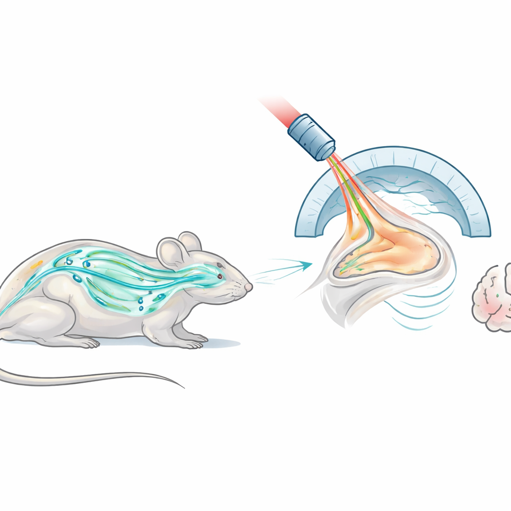
The researchers used an imaging method called photoacoustic computed tomography, or PACT, to monitor CSF movement in live mice. In PACT, short laser flashes gently heat light-absorbing molecules in tissue, causing them to expand and generate ultrasound waves. A curved array of ultrasound detectors then captures these waves and a computer reconstructs three-dimensional images of structures and contrast agents inside the body. To make the otherwise invisible CSF visible, the team injected a medical dye, indocyanine green, into either the spinal fluid or the brain tissue of mice. Because the dye strongly absorbs light at specific colors, PACT could follow where it traveled over minutes to days, while also showing the surrounding anatomy.
Following the Brain’s Cleaning Flow in Real Time
By scanning the whole bodies of mice for 24 hours after dye was delivered into the spinal canal, the team visualized the dye spreading through the fluid-filled space around the spinal cord, reaching a reservoir at the base of the brain called the cisterna magna, and then disappearing as it was cleared. They confirmed the same pattern with a separate fluorescence technique that measures glowing light from the dye. Next, they zoomed in on the brain region and repeatedly imaged it for 30 minutes to see how quickly CSF drained from the cisterna magna under different types of anesthesia. Mice given a common injectable drug mix showed much stronger and faster dye movement than mice inhaling a gas anesthetic, underscoring that brain fluid flow is sensitive to brain state—similar to how previous work has linked deep sleep to more vigorous cleaning.
Signs of Sluggish Cleaning in Alzheimer’s-Like Brains
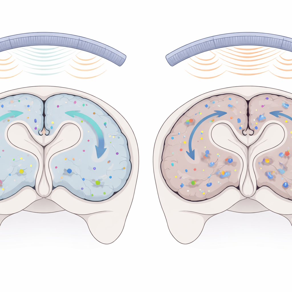
To test whether PACT could detect impaired waste clearance, the scientists turned to a mouse strain that develops amyloid buildup and other features resembling Alzheimer’s disease. This time, they injected a small amount of dye directly into a deep brain region called the striatum and tracked how much remained over four days. In healthy mice, the dye signal faded steadily, indicating ongoing clearance through fluid pathways. In the Alzheimer’s-like mice, however, the dye signal in the same region barely decreased, even after 96 hours, suggesting that waste had trouble leaving the brain tissue. Follow-up measurements on dissected brains using fluorescence imaging confirmed that more dye was retained in the disease-model mice than in their healthy counterparts.
What This Means for Brain Health and Disease
Taken together, the experiments show that PACT can noninvasively track brain fluid movement across the entire body, monitor rapid changes in CSF flow in real time, and reveal long-term differences in how efficiently the brain clears waste. For non-specialists, the key message is that our brains rely on a delicate plumbing and drainage system to stay healthy, and that this system can be measured and compared under different conditions. While this work was done in mice and the method still needs technical refinements, it points toward future tools for studying how aging, anesthesia, and neurological diseases disturb the brain’s self-cleaning abilities—and, eventually, for testing treatments aimed at restoring that vital housekeeping.
Citation: Choi, S., Kim, J., Jeon, H. et al. Photoacoustic computed tomography monitors cerebrospinal fluid dynamics and glymphatic function. Nat Commun 17, 2677 (2026). https://doi.org/10.1038/s41467-026-69390-4
Keywords: cerebrospinal fluid, glymphatic system, photoacoustic imaging, brain waste clearance, Alzheimer’s disease