Clear Sky Science · en
Glycocalyx micro- and nanodomains in cell-cell and cell-matrix interactions revealed by enhanced click chemistry
How Cells Wear a Sugar Coat
Every cell in our body is wrapped in a thin, sugar-rich layer called the glycocalyx. This sugar coat helps cells sense their surroundings, stick to or repel neighbors, and even evade the immune system. In cancer, this coat often becomes thicker and more chaotic, which can encourage tumors to grow and spread. The study summarized here develops a sharper way to see this sugar coat on living cells and reveals that, rather than being a smooth shell, it is full of tiny and small-scale gaps exactly where cells touch each other and the tissue around them.
A New Way to Light Up the Sugar Coat
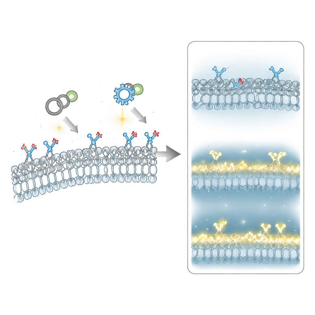
Traditional methods to visualize the glycocalyx rely on antibodies or natural sugar-binding proteins. These tools can tug on the molecules they bind, clump them together, or miss regions where the signal is weak. The authors instead use a two-step “chemical tagging” strategy: cancer cells are first fed a harmless sugar building block that carries a tiny chemical handle. The cells naturally install this altered sugar throughout their surface coat. In the second step, a fluorescent probe snaps onto this handle through a highly selective “click” reaction. The team replaces a widely used click probe (DBCO) with a newer, seven-membered ring probe known as THS, which reacts faster and dissolves better in water. Because THS is more reactive and less sticky to membranes, it labels more of the glycocalyx, gives brighter signals, and keeps background noise low, all without measurably stressing or damaging the cells.
Seeing Finer Details on Living Cells
With this enhanced labeling, combined with high-resolution Airyscan microscopy, the researchers can map the glycocalyx on cancer cells at both micro- and nano-scales while cells are alive. They confirm that their metabolic label tracks most of the major components of the sugar coat, except for the very outer layer of one long-chain sugar (hyaluronic acid). Compared with the older probe, THS reveals a larger fraction of cells with highly labeled coats and improves signal-to-background by several fold, especially on tiny features like membrane blebs. This improvement means that subtle changes in glycocalyx density that were previously too faint or noisy to see now become visible as clear patterns along the cell surface.
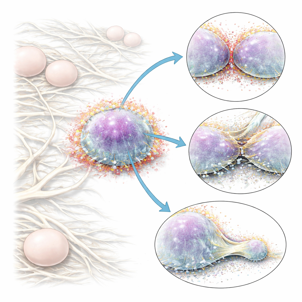
Hidden Gaps Where Cells Touch Each Other and the Matrix
Using these sharper images, the team examines how the sugar coat behaves where cells meet other cells and the surrounding scaffold of fibers known as the extracellular matrix. At first glance, the coat looks fairly even on free cell surfaces. But at cell–cell contacts, the signal is lower than expected if two intact coats simply overlapped, and gentle gradients lead away from these contact zones. Measurements of how quickly fluorescence recovers after bleaching indicate that sugar molecules move faster in these regions, consistent with a dynamic thinning of the coat as cells press together. In three-dimensional collagen gels that mimic soft tissue, cancer cells extend leading-edge protrusions, blebs, and long retraction fibers as they migrate. Along these structures, the glycocalyx progressively thins toward the tips, sometimes over many micrometers, creating low-sugar domains right where the cell pushes into or pulls on the matrix. At even smaller scales, where cells clutch individual collagen fibers, clusters of adhesion proteins called integrins sit slightly outward from a richer inner layer of glycocalyx, forming nanodomains with high integrin and low sugar content side-by-side with more sugar-rich areas.
Why Sugar-Free Patches Matter for Cancer Cells
These observations suggest that cancer cells do not simply carry a uniformly thick sugar coat; they actively sculpt it. By locally thinning the glycocalyx at cell–cell interfaces and at points where they grip collagen fibers, cells may reduce physical crowding around key receptors, making it easier for those receptors to bind partners and transmit mechanical forces. In leading protrusions, reduced sugar density appears to coincide with stronger clustering of integrins, potentially boosting the cell’s ability to latch onto and pull against the surrounding fibers. In blebs and retraction fibers, gradients in the coat align with rapid membrane flow and internal pressure, again pointing to dynamic remodeling rather than a static shell. Overall, the work shows that a more sensitive chemical flashlight—THS-based click chemistry—can uncover micro- and nano-sized “bare spots” in the sugar coat that are likely crucial for how tumor cells communicate, adhere, and move through three-dimensional tissue.
Citation: Smits, D., Damen, J.A.M., Li, T. et al. Glycocalyx micro- and nanodomains in cell-cell and cell-matrix interactions revealed by enhanced click chemistry. Nat Commun 17, 2645 (2026). https://doi.org/10.1038/s41467-026-69242-1
Keywords: glycocalyx, click chemistry, cell migration, cancer cells, cell adhesion