Clear Sky Science · en
An interpretable AI system reduces false-positive MRI diagnoses by stratifying high-risk breast lesions
Smarter Scans, Fewer Unnecessary Procedures
Breast MRI is one of the best tools for finding cancer early, especially in women at high risk, but it often sees “too much,” flagging many harmless spots as suspicious. These gray-area findings lead to anxiety, extra tests, and painful biopsies that ultimately prove benign. This study introduces a new artificial intelligence (AI) system that reads breast MRI scans alongside radiologists and helps distinguish truly dangerous lesions from those that can be safely watched, with the goal of catching cancer without sending so many women to the biopsy table.
The Problem of Uncertain Breast MRI Findings
When radiologists read a breast MRI, they label suspicious areas using a scale called BI-RADS. Category 4 is the most troublesome: it covers lesions with anything from a 2% to 95% chance of being cancer. Because that range is so wide, the current rule of thumb is to biopsy nearly all of them. As a result, many women undergo invasive procedures for lesions that turn out to be benign. On top of that, MRI interpretation is subjective. Different radiologists, especially those with less experience, can disagree about the same scan, sometimes over-calling benign findings or missing subtle cancers. The authors set out to build a tool that reduces this uncertainty while fitting into real-world hospital practice.
An AI Partner Trained on Thousands of Scans
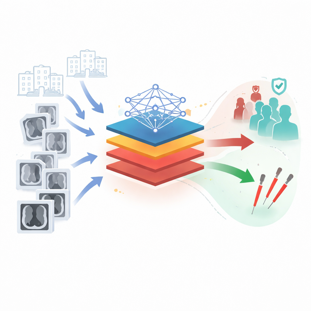
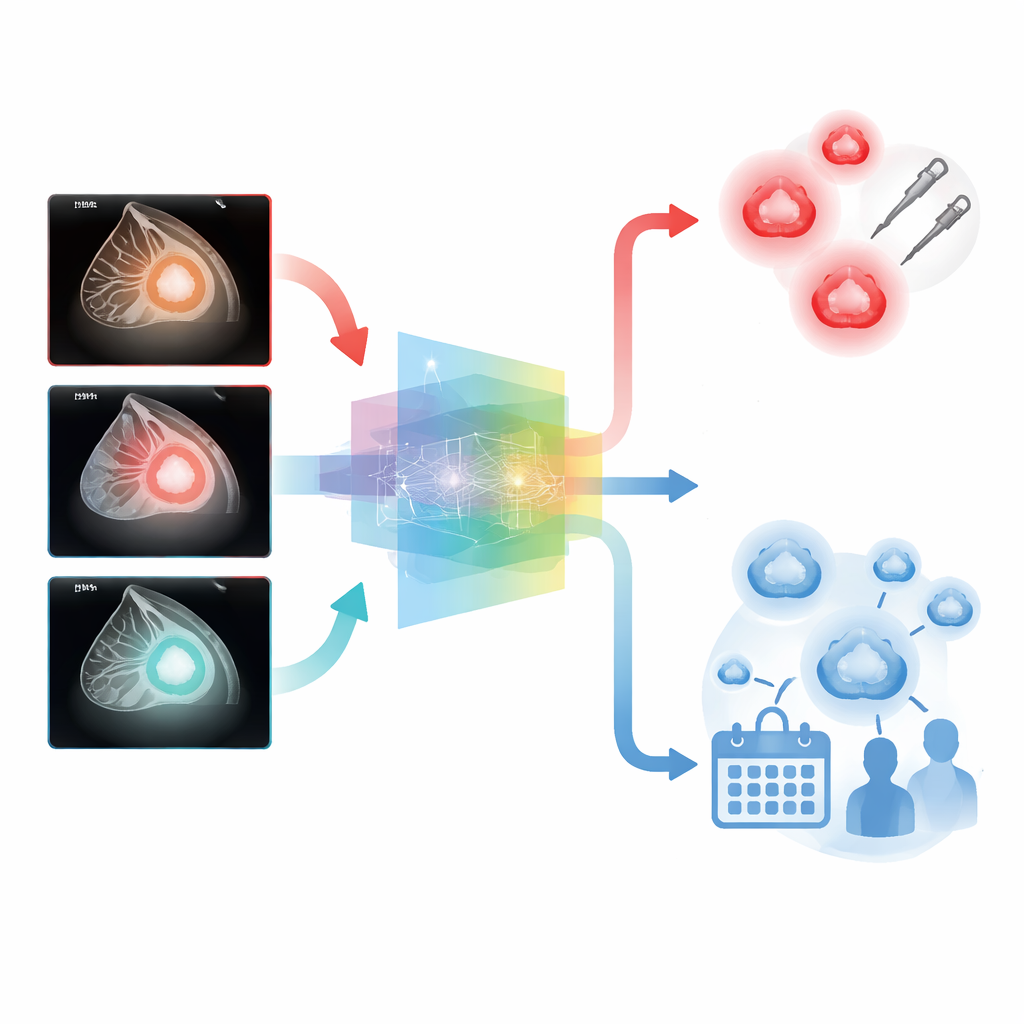
The researchers developed the BI-RADS 4 Lesions Analysis System, or BL4AS, an AI model designed specifically for these high-risk but ambiguous MRI findings. Unlike earlier systems that relied on a single snapshot, BL4AS looks at multiple time points after a contrast dye is injected, tracking how each lesion lights up and fades over time. These changing patterns carry important clues about whether tissue is likely benign or malignant. The team first pre-trained a large “foundation model” on more than 17,000 MRI volumes to learn general imaging features, then fine-tuned it on 2,803 BI-RADS 4 lesions from 2,686 women treated at three medical centers. The system both outlines the lesion and classifies it as low or high risk, offering a probability that it is cancer.
Outperforming Human Readers in Real Clinics
To see how well BL4AS works outside the lab, the authors tested it on independent data from other hospitals and on a fresh, prospectively collected group of patients. Across these settings, the AI showed strong accuracy and, crucially, much higher specificity than radiologists—that is, it was better at recognizing benign lesions and avoiding false alarms. In a prospective reader study, eight radiologists first interpreted cases on their own, then re-read them with BL4AS’s help. With AI support, their diagnostic accuracy rose, their false-positive rate dropped by more than a quarter, and agreement between readers improved markedly. Junior radiologists benefited the most, with their performance nearly matching that of senior colleagues when assisted by the system.
Making AI Decisions Transparent and Actionable
Because clinicians are understandably cautious about “black box” AI, the team built in visual explanations. BL4AS produces heatmaps that highlight which parts of a lesion influenced its decision, often focusing on irregular shapes, sharp edges, and unusual enhancement patterns that radiologists already associate with cancer. The system also goes a step further than a simple yes/no cancer prediction by assigning lesions into BI-RADS 4A, 4B, or 4C subgroups that reflect increasing risk. In external test sets, these AI-defined subcategories closely matched the actual cancer rates and did a better job than radiologists at both identifying low-risk lesions that might safely skip biopsy and flagging high-risk lesions that should be treated promptly.
What This Could Mean for Patients
Overall, the study suggests that an interpretable AI assistant like BL4AS can help radiologists read breast MRI scans more consistently, reduce unnecessary biopsies, and still maintain a very high safety margin against missed cancers. By using the full richness of time-resolved MRI data and presenting its reasoning in a human-friendly way, the system offers a practical path toward more personalized breast cancer care: women with truly worrisome lesions can move quickly to treatment, while those with low-risk findings may avoid invasive procedures and instead be followed closely over time.
Citation: Liang, Y., Wei, Z., Dai, Y. et al. An interpretable AI system reduces false-positive MRI diagnoses by stratifying high-risk breast lesions. Nat Commun 17, 2263 (2026). https://doi.org/10.1038/s41467-026-69212-7
Keywords: breast MRI, artificial intelligence, cancer diagnosis, medical imaging, risk stratification