Clear Sky Science · en
Miniature endoscope for high resolution electrophysiological recordings from the colon of live mice
Why this tiny camera matters for gut health
Many digestive disorders, from chronic constipation to irritable bowel syndrome, are driven by hidden electrical signals that coordinate how the gut squeezes and moves its contents. Until now, scientists studying these signals in small animals have had to rely on crude timing tests or invasive surgery that offer only a blurry picture of what is really happening. This paper introduces a pencil-thin endoscope that can slip into the colon of a live mouse and listen in on thousands of tiny electrical bursts with unprecedented detail, opening the door to faster, more precise discoveries about gut diseases and potential treatments. 
A new window into the working gut
The colon is lined with muscle and its own built-in "brain," the enteric nervous system, which together generate electrical pulses that drive waves of contraction. Traditional tools can measure how long it takes material to pass through the gut or record from just a few spots at a time, missing how activity is coordinated over distance. The researchers set out to build a device that could capture these electrical patterns along a stretch of bowel in a live animal, without cutting the abdomen open or stitching electrodes onto the outer surface.
How the miniature endoscope works
The team designed a semi-rigid tube about 2 millimeters wide and 3 centimeters long—similar in size to a mouse stool pellet—wrapped in a thin flexible film carrying 128 tiny metal sensors. These sensors, coated to reduce electrical resistance, sit directly against the moist inner lining of the colon once the device is gently inserted through the rectum under anesthesia. Bench tests in salty solution and measurements inside the mouse showed that the sensors kept good contact with tissue and could each detect local signals rather than a smeared average, thanks to their small size and careful spacing. Together, the array provides a high‑resolution map of electrical activity along and around the colon wall.
Listening to the colon in action
Using this endoscope in healthy mice, the scientists recorded sharp electrical spikes produced by smooth muscle cells. These spikes grouped into repeating patterns: short bursts roughly twice per minute that traveled along the colon, and faster "burstlets" within each burst about once per second. The device could distinguish waves moving toward the anus from those moving backward, and revealed additional rhythms that were hard to see by eye but emerged when the team analyzed the strength of the signals over time. 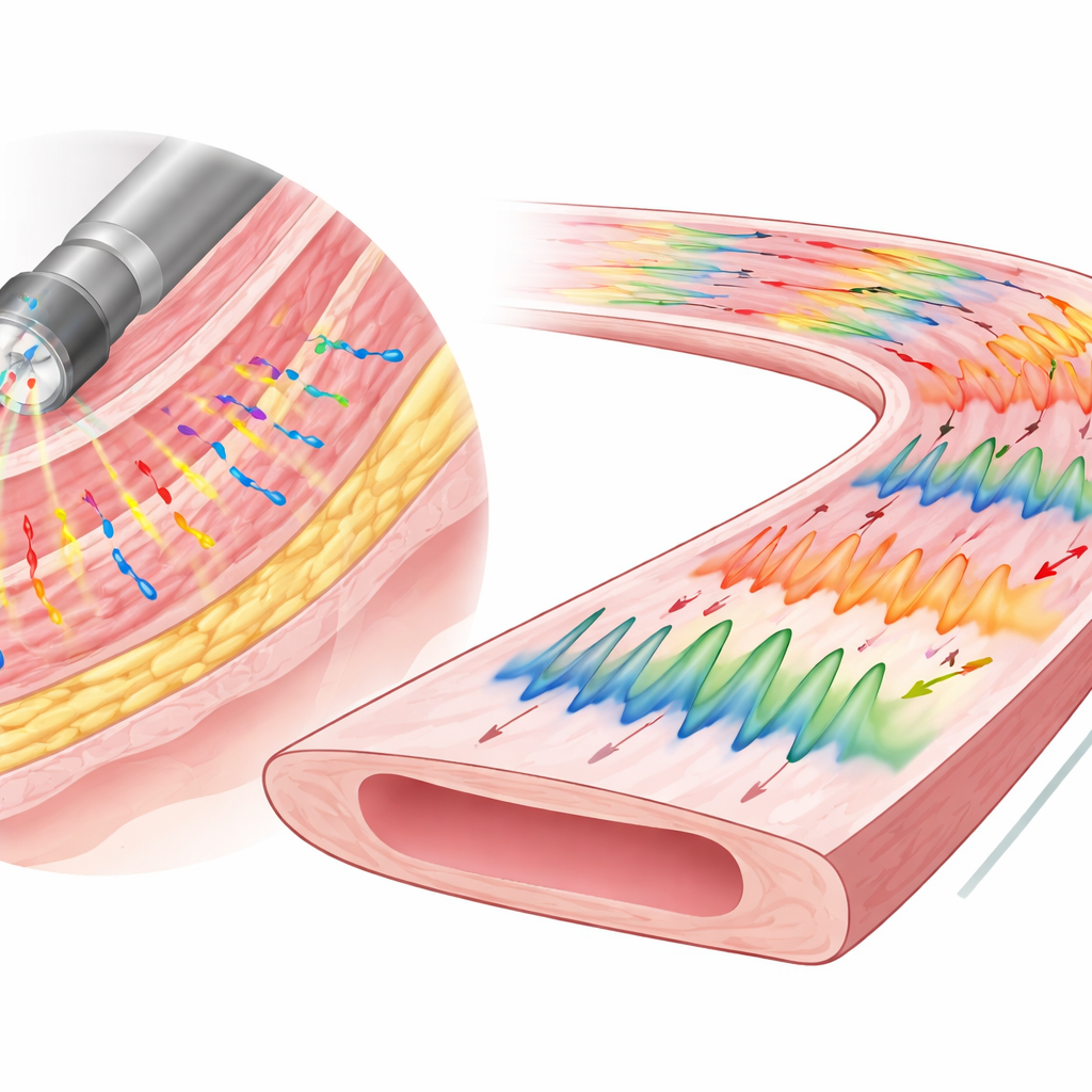
Probing drugs and disease in real time
Because the method is minimally invasive and fast to set up, the researchers could watch how the colon’s electrical behavior changed as they altered its chemistry. A drug that boosts the action of the nerve messenger acetylcholine quickly increased spiking, while a blocker of the same messenger dampened activity, especially in regions that normally show strong rhythmic bursts. In mice whose colons had been chemically injured to disrupt their internal nerve network, the usual regular patterns disappeared and were replaced by irregular, animal‑specific signatures—electrical analogues of arrhythmias. In a separate set of experiments on excised colons kept alive in a warm bath, the endoscope’s recordings matched those from a standard suction electrode and lined up closely with visible contractions captured on video. Blocking nerve signals or calcium entry into muscle cells reshaped or abolished the spikes, confirming that the device was truly measuring the gut’s own control system.
What this means for future gut research
This miniature endoscope turns the mouse colon into an accessible testbed where researchers can directly see how electrical waves change with genes, injury, or candidate drugs, without major surgery. By mapping activity at high resolution across a meaningful length of bowel, it bridges the gap between simple transit tests and complex imaging, and could help explain why some guts push too slowly, too quickly, or in the wrong direction. Ultimately, tools like this may speed the path from basic discoveries about the gut’s "second brain" to targeted therapies for human digestive disorders.
Citation: Sobolewski, A., Planchette, A., Wójcicki, K. et al. Miniature endoscope for high resolution electrophysiological recordings from the colon of live mice. Nat Commun 17, 2363 (2026). https://doi.org/10.1038/s41467-026-69144-2
Keywords: colon motility, enteric nervous system, electrophysiology, mouse model, gut endoscope