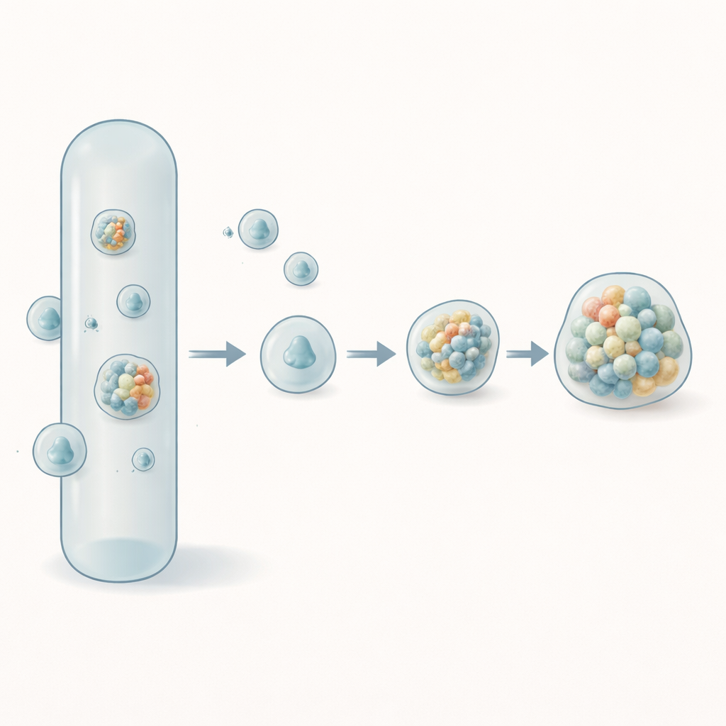
Inside our cells, many crucial reactions take place not in rigid compartments, but in soft, droplet-like pockets made of proteins and other molecules. These droplets can help organize the cell—but they can also go wrong and turn into the harmful clumps seen in neurodegenerative diseases. This study focuses on an especially important protein linked to such diseases and shows, for the first time in fine detail, how this protein forms extremely small droplets—"nanocondensates"—long before visible clumps appear.
Tiny droplets in a crowded cell
Cells are packed with molecules jostling for space, and one way they stay organized is by forming tiny liquid droplets without using membranes. These droplets, called biomolecular condensates, help control gene activity, build cellular machines, and respond to stress. The protein examined here is TDP-43, which plays roles in processing RNA and is strongly associated with conditions such as amyotrophic lateral sclerosis (ALS) and frontotemporal dementia. The authors focus on a floppy tail region of TDP-43, known for driving both droplet formation and the buildup of disease-related aggregates. Understanding how this region first comes together at very small scales could reveal how healthy organization tips into harmful aggregation.
Watching single droplets one by one Figure 1.
To probe these earliest steps, the researchers built a highly sensitive fluorescence setup on a confocal microscope. They labeled a small fraction of TDP-43 molecules with a dye and shone a laser into a tiny observation spot in solution. As individual protein clusters drifted through this spot, they produced brief flashes of light. Instead of averaging all signals, as traditional methods do, the team analyzed every flash separately—its brightness, how long it lasted, and how often such events occurred. This allowed them to count and characterize individual nanocondensates between about 40 and 400 nanometers in size, whose existence is invisible to standard microscopy.
Triggering and mapping the birth of nanocondensates
The team then explored how changing conditions alters droplet formation. They used a small molecule, TMAO, that compacts flexible proteins and encourages them to come together, and they varied both TDP-43 and TMAO concentrations. They found that nanocondensates formed quickly—within about a minute—and at protein levels roughly ten times lower than those needed to see microscopic droplets by eye. By counting events and measuring their total brightness, they built a "phase map" showing where in this concentration space nanocondensates appear. They also repeated the experiments in a cell-like extract, which contains many other biomolecules, and observed similar trends: TDP-43 still formed nanocondensates rapidly, suggesting that this behavior is an intrinsic feature of the protein rather than an artifact of a simple buffer.
How droplets grow, merge, and change over time Figure 2.
Because each flash of light could be fingerprinted by its intensity and duration, the researchers could follow how droplet properties evolved. Larger, slower-moving droplets produced wider peaks, allowing the team to estimate physical size using both simulations and calibration beads. Most TDP-43 nanocondensates were about 100–250 nanometers across, and their size depended more on protein concentration than on TMAO level. Over tens of minutes, many small, fast-diffusing condensates gradually gave way to fewer, larger ones, consistent with droplets merging or growing. When the team mixed green- and red-labeled droplets, they saw the colors blend over time, showing that material exchanges between condensates and that they behave like liquids rather than rigid particles. A chemical that weakens hydrophobic interactions could dissolve most droplets, further reinforcing their liquid-like nature.
From soft droplets to harmful aggregates
Nanocondensates are not necessarily permanent or benign. TDP-43 is notorious for forming amyloid-like fibrils in disease, so the authors asked whether some droplets eventually harden into more solid structures. Using a dye that glows when it binds amyloid, they tracked both droplets and emerging aggregates in two colors at once. Early on, droplets were dye-negative, but after hours—or sooner at higher protein levels—a subset of slowly moving, larger condensates became dye-positive, signaling amyloid content. Crucially, only a fraction of droplets followed this path; many remained liquid-like and dye-negative, highlighting that not all condensates are equally prone to becoming harmful aggregates.
What this means for brain disease and beyond
This work shows that disease-linked proteins like TDP-43 begin organizing into nanoscopic droplets at much lower concentrations and much earlier times than previously appreciated. By following individual droplets, the method distinguishes between reversible liquid organization and the later emergence of more solid, amyloid-containing structures. For a layperson, the key message is that before large, visible clumps appear in conditions like ALS, there is an invisible world of tiny droplets that may set the stage for disease. The single-droplet toolkit demonstrated here offers a powerful way to study that hidden world and could ultimately guide strategies to nudge proteins back toward healthy liquid behavior and away from damaging solid aggregates.
Citation: Houx, J., Cussac, J., Copie, T. et al. Direct observation and quantification of single nanocondensates of the low complexity domain of TDP-43.
Nat Commun17, 2505 (2026). https://doi.org/10.1038/s41467-026-69024-9
Keywords: protein droplets, TDP-43, nanocondensates, phase separation, neurodegeneration