Clear Sky Science · en
Automated diagnostic of cervical spondylosis on multimodal medical images with a multi-task deep learning model
A Hidden Neck Problem With a Big Everyday Impact
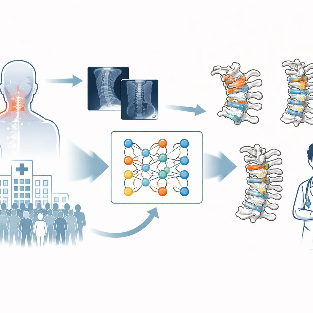
Cervical spondylosis—age- or lifestyle-related wear and tear in the neck—is nearly everywhere, yet often hard to spot early. It can cause nagging neck pain, headaches, numbness, and even trouble walking, but its subtle changes on medical images demand years of experience to read. This study shows how an artificial intelligence (AI) system can learn from expert doctors to read X‑rays and MRI scans of the neck, helping bring specialist‑level diagnosis to busy hospitals and under‑served clinics alike.
Why Neck Wear and Tear Is So Hard to Catch
Unlike a broken bone or a large tumor, cervical spondylosis usually does not leave a single obvious mark on a scan. Instead, it involves many small, interacting changes: the natural curve of the neck may flatten or reverse, vertebrae can shift slightly out of place, discs may bulge or rupture, and the canal that houses the spinal cord can slowly narrow. Doctors currently measure several geometric features on X‑rays—such as the overall bend of the neck and the alignment of key vertebrae—as well as changes seen on MRI that reveal soft‑tissue problems like disc herniation or pinched nerve roots. Doing this accurately for every patient is time‑consuming and depends heavily on the skill of seasoned radiologists and spine surgeons, who are often in short supply outside major medical centers.
Teaching a Computer to Read the Neck Like a Specialist
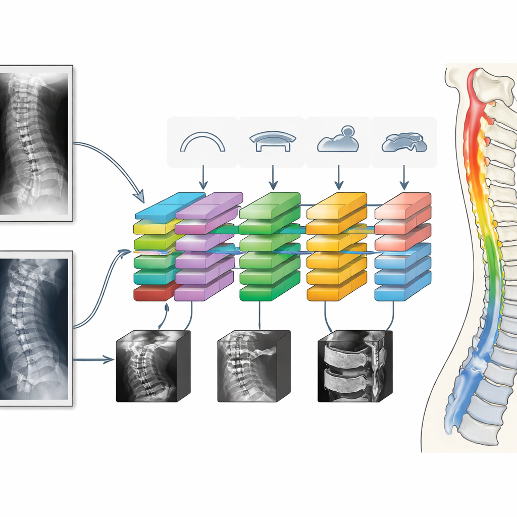
The researchers built a deep learning system that mimics how experts look at cervical images, but does so automatically and consistently. First, it detects the outlines and corners of the neck vertebrae on lateral X‑rays, treating each vertebra as a precisely defined quadrilateral. From these points, the model computes standard measurements used in surgery planning, such as the Cobb angle (which describes how curved or straight the neck is) and the sagittal vertical axis, a key marker of whether the head is well balanced over the spine. It also estimates how much neighboring vertebrae slip relative to each other, signaling instability, and measures the size of the spinal canal and vertebral body to calculate a ratio that indicates possible congenital narrowing.
Combining Different Scans Into One Coherent Picture
Because no single scan type tells the whole story, the team designed the system to work with both X‑rays and MRI in a coordinated way. X‑rays provide precise information about bone shape and alignment, while MRI shows the condition of soft tissues, especially the intervertebral discs and the openings where nerves exit the spine. Using a multi‑task learning setup, the AI is trained to perform all these measurements and classifications together, sharing what it learns between tasks. For MRI, a dedicated network looks at the discs between each pair of vertebrae and determines whether they are relatively healthy or show more serious bulging or extrusion, and whether the spinal canal and nerve exits are narrowed. Instead of fusing everything into a single yes‑or‑no label, the system outputs a detailed profile of which problems are present and where.
How Well the AI Stacks Up Against Human Doctors
The study analyzed X‑rays and MRI scans from over a thousand patients treated at a major hospital, with careful labels provided by experienced radiologists and spine specialists. On simple geometric measures like distances, the AI’s errors were often under a millimeter, and on angles they were only a few degrees off on average—close enough to be considered clinically interchangeable with manual measurements in many cases. For disc problems, canal narrowing, and nerve exit stenosis on MRI, the model reached accuracy levels comparable to, and sometimes better than, junior and senior radiologists, and similar to seasoned clinicians, while producing its results in seconds rather than minutes. When the authors tested the system at a second hospital, the performance dipped slightly but remained high, suggesting it can generalize beyond the original training site.
What This Could Mean for Patients and Clinics
To a person living with neck pain, the promise of this work lies in faster, more consistent diagnoses and better‑tailored treatment decisions. The AI does not replace doctors; instead, it serves as a tireless assistant that can pre‑measure key features, flag likely problem areas, and provide a unified, scan‑by‑scan report of neck health. Because it mirrors the measurements experts already trust—rather than inventing new ones—it can slot into existing clinical routines and help transfer specialist knowledge to hospitals that lack in‑house spine experts. If adopted widely and validated further, such systems could lead to earlier detection of risky neck changes, fewer missed cases, and more appropriate surgery or conservative care for millions of people worldwide.
Citation: Song, X., Li, Y., Ouyang, H. et al. Automated diagnostic of cervical spondylosis on multimodal medical images with a multi-task deep learning model. Nat Commun 17, 2392 (2026). https://doi.org/10.1038/s41467-026-69023-w
Keywords: cervical spondylosis, spine imaging, deep learning, medical AI, neck pain