Clear Sky Science · en
A spatiotemporal atlas of cerebrovascular development in zebrafish
Why tiny fish brains matter
The brain is one of the body’s thirstiest organs, but its blood vessels must do more than just deliver oxygen. They also form a protective filter called the blood–brain barrier, which lets in nutrients while blocking toxins. When this vascular system goes wrong, the result can be strokes, dementia, or other neurological diseases. This study uses transparent zebrafish larvae to watch, in three dimensions and at single-cell resolution, how brain blood vessels grow and how the brain’s protective barrier switches on during early life.
Building a living road map of brain vessels
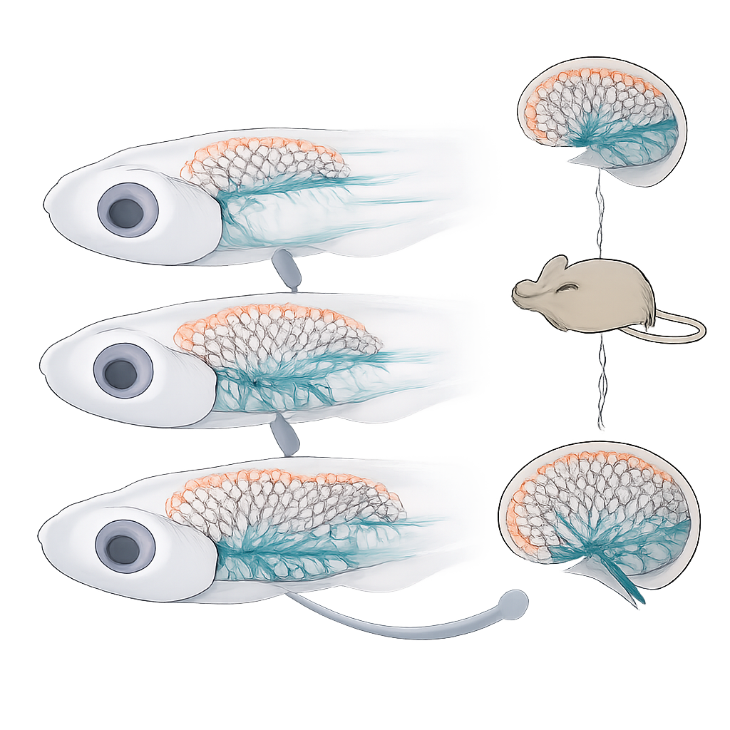
The researchers first created a three-dimensional atlas of blood vessels in the zebrafish brain from three to eleven days after fertilization. Using a fluorescent reporter that lights up the cells lining blood vessels, they reconstructed the entire brain vasculature at each stage. Their measurements showed that total vessel length and the number of segments expand dramatically over this short window. Early on, most new vessels sprout along the sides of the brain. As development proceeds, growth shifts inward, with a burst of small vessels penetrating deep into brain tissue. This pattern marks a transition from a simple outer scaffold of vessels to a dense internal network that directly serves brain cells.
Finding the main players in vessel walls
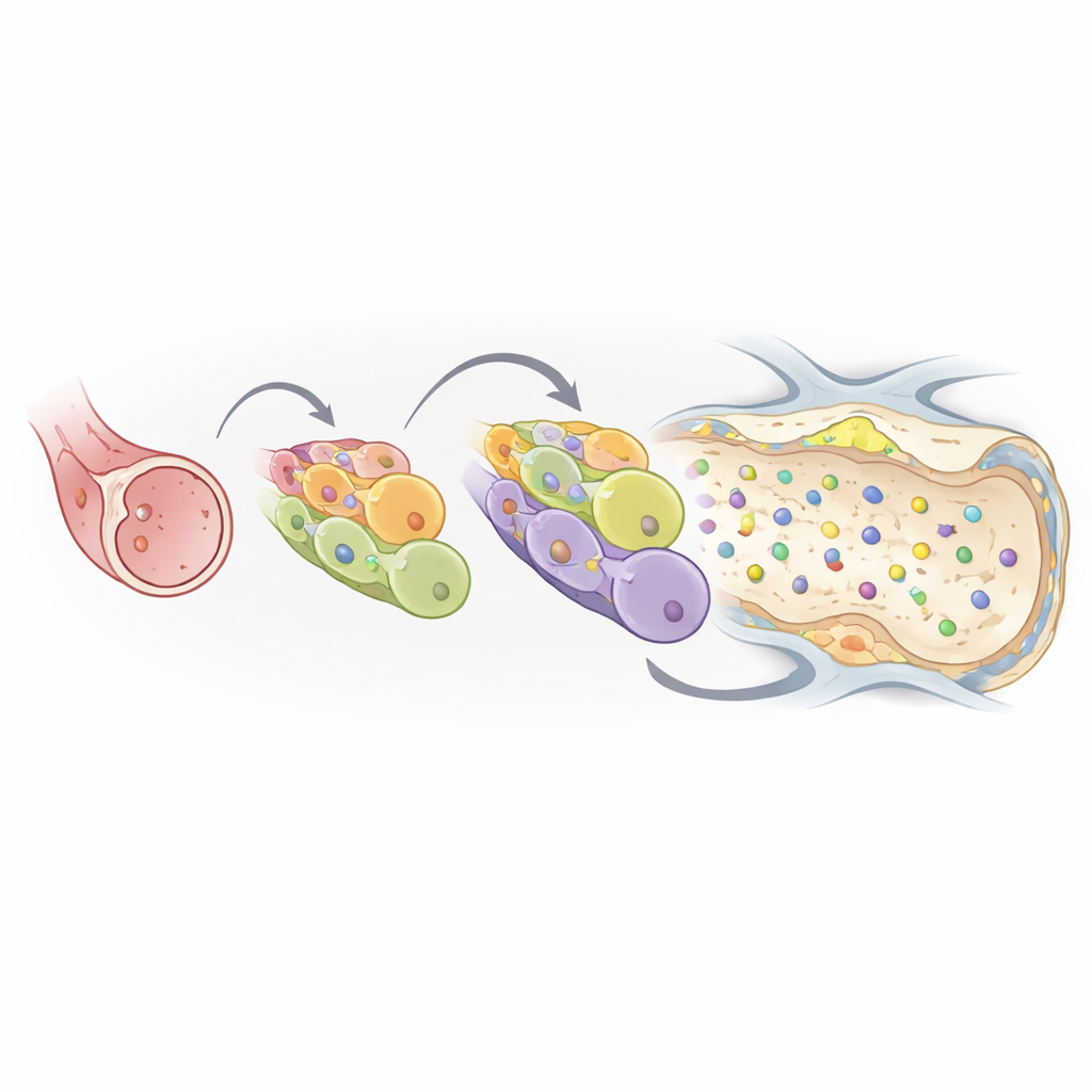
Brain blood vessels are lined by endothelial cells, but not all endothelial cells are the same. To understand who does what, the team isolated these cells from zebrafish brains at each stage and profiled their gene activity one cell at a time. They identified six major endothelial subtypes, including arterial, venous, lymphatic, actively dividing, sprouting, and capillary cells. Capillary endothelial cells emerged as the dominant type in the intracranial network and showed strong enrichment of genes involved in pumping substances across the vessel wall and sealing the spaces between neighboring cells. These features are key hallmarks of a functional blood–brain barrier.
Pinpointing when the brain’s barrier closes
To connect molecular fingerprints with location, the researchers overlaid single-cell data onto spatial maps of the whole brain. Using an in situ sequencing method, they recorded where dozens of marker genes were expressed in thin brain sections, then aligned those sections back onto the three-dimensional vascular map. This revealed that capillary-like endothelial cells gradually accumulate in midbrain and hindbrain vessels, while arterial cells become concentrated in forebrain arteries. Parallel experiments tracking dye leakage from the bloodstream showed that the barrier is leaky at three and six days, but by eleven days the dye remains confined inside brain vessels. Gene modules for transporters and tight junction components ramp up over time in capillary cells, matching the observed closure of the barrier.
Shared patterns from fish to humans
The team then asked whether these zebrafish vessel types resemble those in mammals. By comparing their zebrafish data with published single-cell profiles from developing mouse and human brains, they found strong conservation of endothelial subtypes and of key gene modules, especially in capillary cells. This suggests that zebrafish provide a faithful stand-in for early human cerebrovascular development. From their comprehensive dataset, the authors highlighted three capillary-enriched genes that had not been functionally tested in this context. Using genome editing and gene knockdown, they showed that two transporter genes and one barrier-associated gene are needed for proper vessel growth and stable brain vasculature; disrupting them alters vessel patterning and can cause bleeding or a leaky barrier.
What this means for brain health
Taken together, the work delivers a multidimensional atlas that links vessel architecture, cell types, and gene activity across early brain development in a living vertebrate. For non-specialists, the key message is that the tiny zebrafish brain recapitulates many essential features of human brain vasculature, including the stepwise tightening of the blood–brain barrier and the specialization of different vessel segments. This atlas and the newly identified barrier genes offer a framework for probing how brain vessels form, how they fail in disease, and how they might be targeted to deliver therapies more effectively into the brain.
Citation: Li, X., Ke, S., Wu, C. et al. A spatiotemporal atlas of cerebrovascular development in zebrafish. Nat Commun 17, 2216 (2026). https://doi.org/10.1038/s41467-026-68995-z
Keywords: blood-brain barrier, zebrafish, brain vasculature, endothelial cells, single-cell transcriptomics