Clear Sky Science · en
Cryogenic transmission electron microscopy reveals assembly and nanostructure of PEDOT:PSS
Stretchy Wires You Can Wear
Imagine electronics so soft and stretchy that they can bend with your skin, wrap around a beating heart, or move with your muscles without breaking. A plastic-like material called PEDOT:PSS is already at the heart of many such bioelectronic and wearable devices. Yet, until now, scientists have not had a clear picture of how its tiniest building blocks arrange themselves, or why certain recipes make it both highly conductive and remarkably stretchable. This study uses powerful electron microscopes at ultra‑cold temperatures to watch PEDOT:PSS assemble from solution into solid films, revealing how small structural changes unlock big performance gains.
Looking Closer at a Workhorse Material
PEDOT:PSS is a mix of two polymers: one that carries electrical charges and one that helps it dissolve in water and form films. By itself, this mixture conducts electricity only modestly well and is not very tough when stretched. Manufacturers have learned that adding certain salts or small molecules can boost conductivity by up to a thousand-fold and make the films more flexible, but the microscopic reasons for this behavior were murky. Traditional tools like X‑ray and neutron scattering hinted at structures inside the material, yet they could not directly show how those structures looked in real space, especially in the wet environments where many devices actually operate.
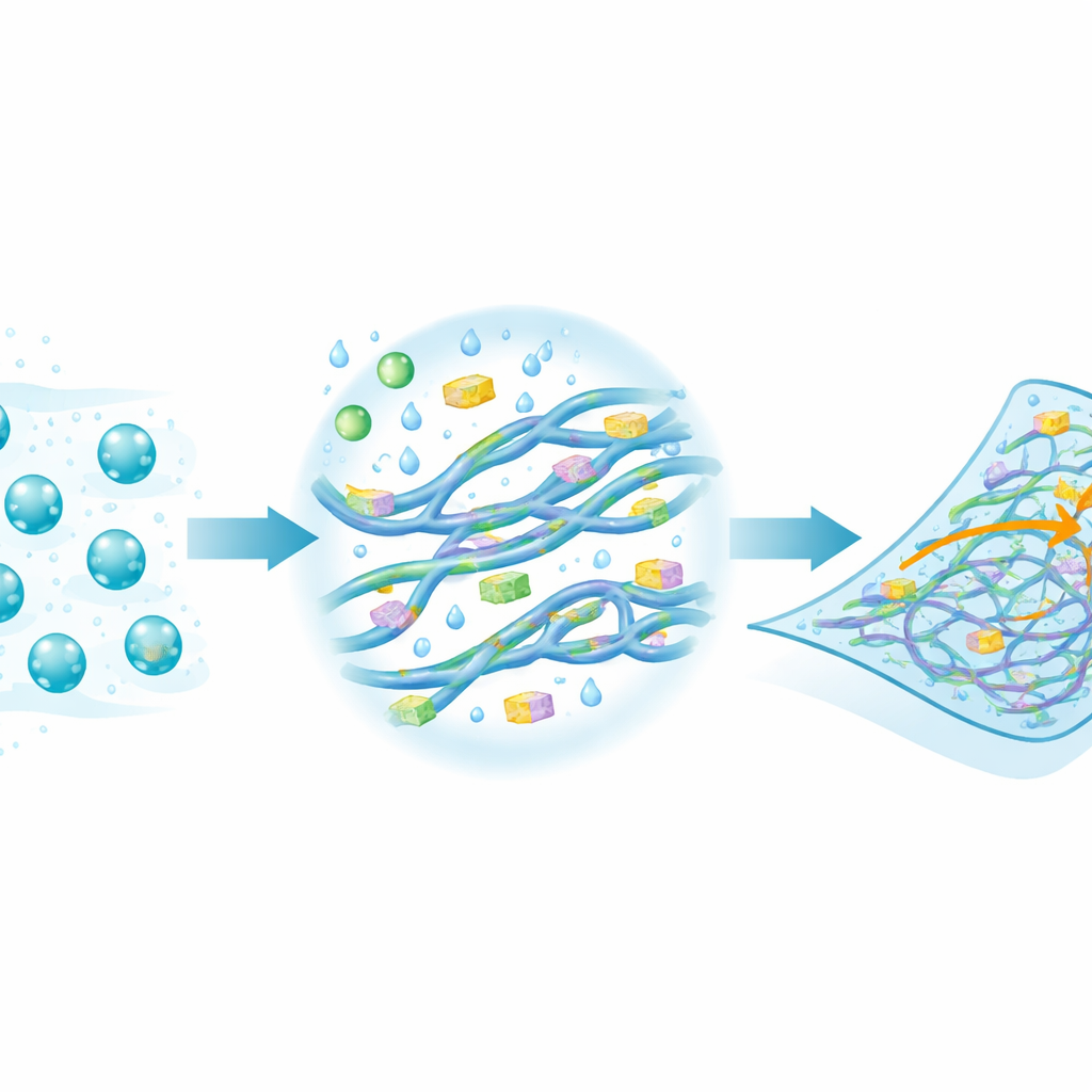
Freezing Motion to Reveal Hidden Shapes
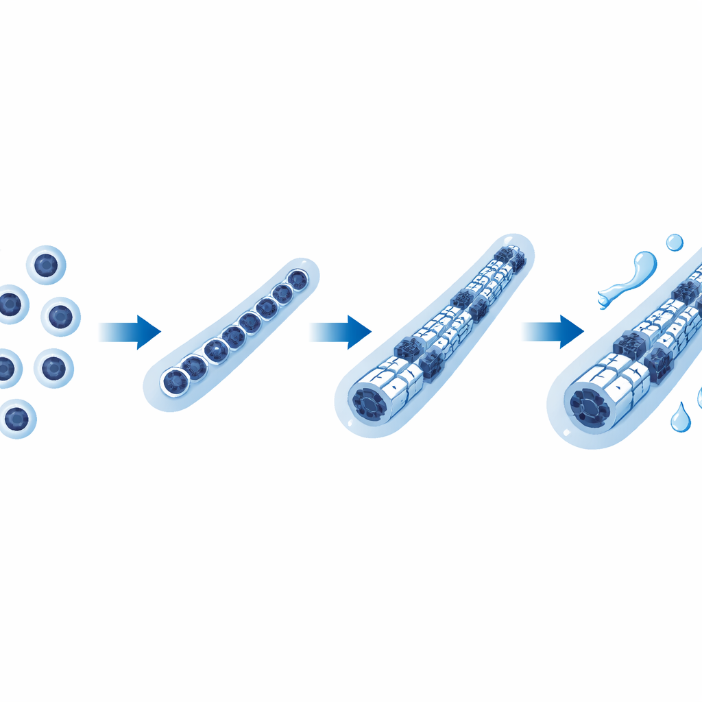
The researchers turned to cryogenic transmission electron microscopy, or cryo‑EM, a technique that flash‑freezes liquid samples so fast that their internal structure is preserved in place. Starting from PEDOT:PSS in water, they saw tiny spherical clusters known as micelles, along with a few thin, elongated fibers. When they added ionic salts or a non‑ionic additive used in soft electronics, these fibers became much more abundant and were wrapped by short, regularly spaced polymer stacks that signal emerging crystalline order. The images show that fibers form when many micelles merge and their chains begin to stack side‑by‑side, building what the authors call heterostructural fibers—complex strands combining mixed regions and more ordered patches.
From Liquid Threads to Solid Films
Next, the team examined thin solid films made from these solutions. In additive‑free films, they found small crystalline regions and micelles but could no longer clearly see the elongated fibers, suggesting that the few fibers present in solution had merged or broken up. In contrast, films made with salts or other additives contained a rich landscape: long fibrils built from coalesced micelles and numerous crystalline domains, some over 20 nanometers across. This close match between structures in liquid and in solid form shows that what happens in solution—the growth of fibers and nascent crystals—templates the architecture of the final film. X‑ray scattering measurements backed up these images, confirming the presence of both mixed polymer stacks and regions dominated by the conductive component.
Water as a Hidden Design Partner
Because many PEDOT:PSS devices operate in contact with sweat, tissue, or other liquids, the authors also probed what happens when the films take up water. Using cryo‑EM on hydrated films and automated image analysis software, they discovered a striking contrast: the elongated fibers swell noticeably as water penetrates their softer outer layers, while the crystalline regions shrink into smaller domains. At the same time, measurements of stretching behavior showed that films containing additives withstand much larger strains when humid than when dry, and thermogravimetric tests and elemental mapping revealed that additives encourage the material to absorb more water. Together, these results suggest that salts and similar molecules act like built‑in water attractors, forming water–salt complexes that soften parts of the polymer network without destroying its conducting pathways.
Why This Matters for Future Wearable Tech
Putting these pieces together, the study paints a new picture of how PEDOT:PSS can be both highly conductive and mechanically forgiving. Additives help micelles merge into a connected fiber network and promote crystalline regions that carry charge efficiently. When the material hydrates, the fibers swell and the surrounding polymer becomes softer, creating a stretchable scaffold, while the smaller but plentiful crystalline pockets maintain electrical performance. Instead of a simple trade‑off between stiffness and conductivity, PEDOT:PSS can, with the right additives and moisture, behave like a flexible metallic mesh embedded in a soft gel. This deeper structural understanding offers a roadmap for designing next‑generation mixed‑conductor polymers for uses ranging from implantable electrodes and soft sensors to brain‑inspired computing devices.
Citation: Ghasemi, M., Kirkley, L.Y., Nazari, F. et al. Cryogenic transmission electron microscopy reveals assembly and nanostructure of PEDOT:PSS. Nat Commun 17, 2555 (2026). https://doi.org/10.1038/s41467-026-68890-7
Keywords: PEDOT:PSS, cryo-EM, stretchable electronics, mixed ionic-electronic conductors, bioelectronics