Clear Sky Science · en
Directed cortico-limbic dialogue in the human brain
Why this brain story matters
Every thought, feeling, or memory you have depends on signals racing through a vast web of nerve cells. Yet even today, scientists still struggle to see which brain areas actually send information and which ones mostly listen. This study takes advantage of a rare clinical opportunity in people with epilepsy to directly poke the brain with tiny electrical pulses and watch how signals travel. By doing so over many days and during both wakefulness and sleep, the researchers challenge long-held ideas about how emotional and memory centers talk with the rest of the brain.
A rare window inside the living human brain
When people with severe epilepsy are evaluated for surgery, doctors sometimes implant fine electrodes deep in the brain to find where seizures start. The research team used these same electrodes for an additional purpose: instead of only listening, they also briefly stimulated one spot and measured how other areas responded. Each pulse was like tapping one node in a network and seeing which other nodes blinked back. By repeating this procedure hundreds of times per connection, across 15 volunteers and over many hours of hospital monitoring, the team assembled more than three million cause-and-effect measurements of signal flow between brain regions.
Mapping the brain’s talking neighborhoods
To make sense of this flood of data, the researchers grouped tiny recording sites into larger, functionally connected regions. These included the outer thinking layer of the brain (the neocortex) and deeper “limbic” structures such as the hippocampus and amygdala, which are crucial for memory and emotion. For each pair of regions, they asked two basic questions: how often does a signal successfully travel along this pathway when we stimulate it, and does the traffic prefer one direction over the other? Instead of averaging responses into a single trace, they looked trial by trial, revealing that some connections behaved like reliable highways while others were more like side streets that only occasionally carried a pulse through.
Who speaks and who listens?
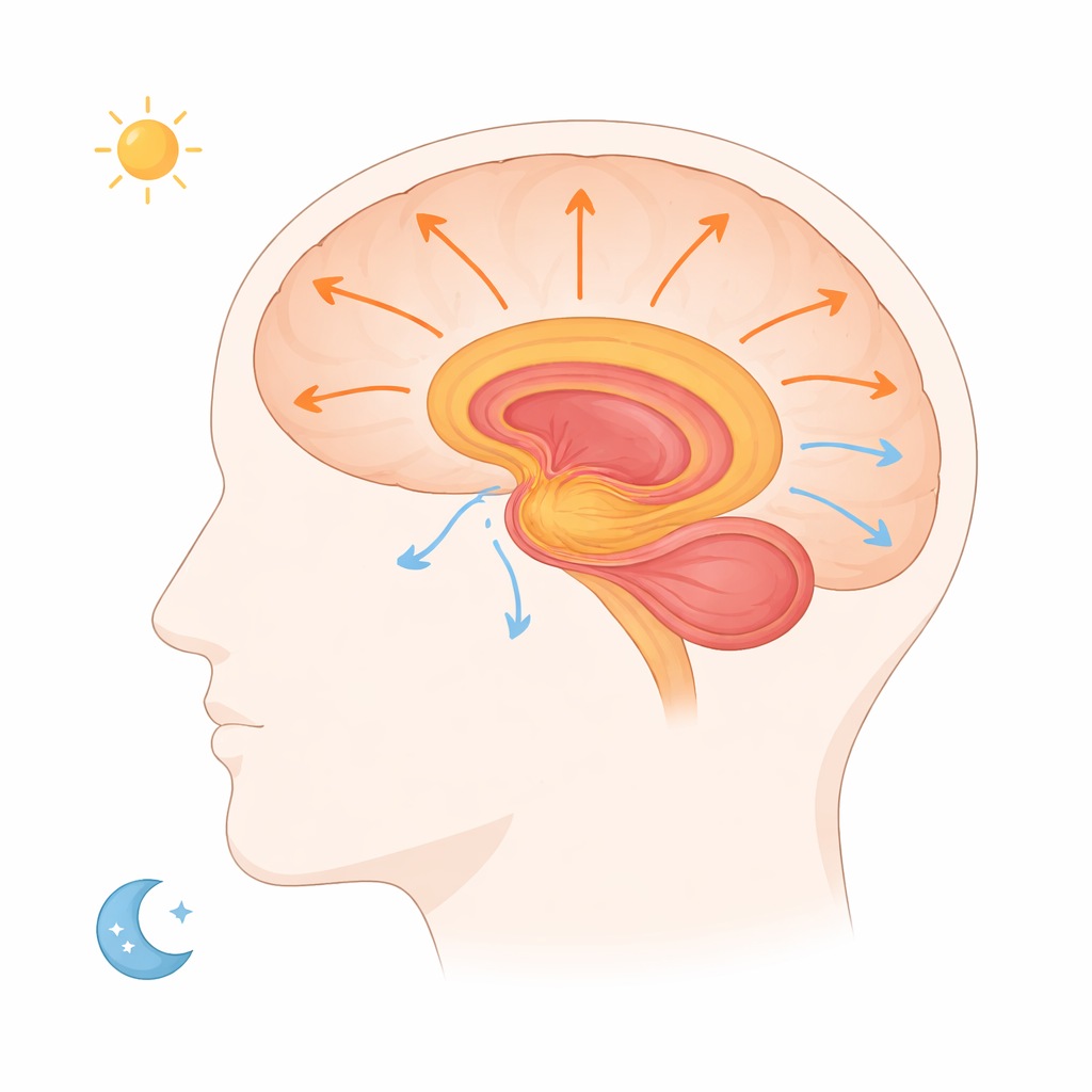
Close-by areas within the same neighborhood of cortex almost always responded strongly and in both directions, suggesting dense two-way chatter over short distances. Long-range links told a different story. Signals between far-apart regions were less dependable and often strongly biased in one direction. Contrary to the traditional picture where the neocortex is seen as the main driver and the limbic system as a receiver, the data showed that limbic structures tended to send about twice as many signals as they received. The amygdala and hippocampus in particular broadcast robust outputs toward frontal and cingulate areas involved in decision making and emotional evaluation. How often a pathway carried a signal closely matched how easily it could be activated, an “excitability” property that varied from connection to connection.
What happens when the brain sleeps
For decades, a popular theory has held that during waking life information flows from the senses into limbic memory systems, while during sleep the direction reverses as memories are “replayed” back to the cortex for long-term storage. Because this study could stimulate the same pathways during wakefulness and different sleep stages, it provided a rare causal test of this idea. The overall pattern of which regions could talk to which stayed remarkably stable between wake and sleep. Some connections even became slightly more excitable at night. But rather than a sweeping flip in direction, the researchers saw something subtler: key outputs from the hippocampus to frontal and cingulate regions actually became weaker and less frequent during both deep and dreaming sleep.
Rethinking brain conversations
This work suggests that, in humans, the limbic system acts less like a passive inbox and more like an active broadcaster, sending information outward across the brain both when we are awake and when we are asleep. The expected wholesale reversal of traffic during sleep did not appear; instead, specific memory pathways quieted down while others changed only modestly. Beyond overturning a classic view, the study offers a new, openly available map of directed brain communication built from direct cause-and-effect tests. In the long run, understanding which connections normally lead and which follow could help doctors design more precise electrical therapies for brain disorders where these conversations go awry.
Citation: van Maren, E., Mignardot, C.G., Widmer, R. et al. Directed cortico-limbic dialogue in the human brain. Nat Commun 17, 2258 (2026). https://doi.org/10.1038/s41467-026-68701-z
Keywords: brain connectivity, limbic system, memory and emotion, sleep and wakefulness, intracranial stimulation