Clear Sky Science · en
Superelastic Tellurium Thermoelectric Coatings for Advanced Trimodal Microsensing
Why adding “touch” to tiny cameras matters
Doctors increasingly rely on endoscopes—thin, flexible cameras—to look inside the body without major surgery. These tools can already show color images and sometimes measure how hard they are pressing on tissue. But they still miss a vital clue: temperature. Many diseases, including inflammation and some tumors, make tissue slightly warmer or stiffer than normal. This study introduces a new type of endoscope tip that can see, feel, and sense heat all at once, potentially helping doctors spot hidden problems earlier and more safely.
A soft window into the body
The researchers built a tiny sensing cap, just a few millimeters across, that can be attached to the front of a standard medical endoscope. The cap is made from a clear, rubbery silicone so light from the camera can still pass through. Hidden within this soft dome are microscopic patterns made from a special material based on the element tellurium. These patterns act like tiny landmarks. When the dome presses on tissue, the landmarks move in subtle ways that a camera can see, letting computers work out how hard and in which direction the probe is pressing. At the same time, the tellurium patterns act as contact thermometers, turning small temperature differences into electrical signals without blocking the doctor’s view. 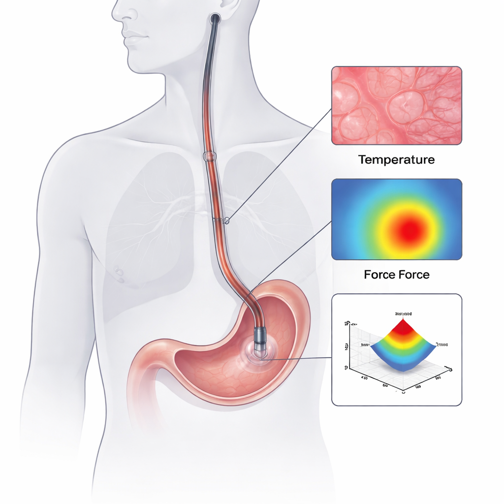
Turning heat into helpful signals
The key to temperature sensing lies in the tellurium coating. Tellurium’s crystal structure naturally hampers heat flow, so when one side touches warmer tissue and the other faces cooler surroundings, a sharp temperature difference builds up across the thin film. This gradient produces a tiny voltage—like a miniature power cell—that rises steadily with temperature. The team engineered the coating to be only about 200 nanometers thick and less than a square millimeter in area, yet it still generated clear, stable signals. Tests showed that the voltage changed in a nearly straight-line fashion with temperature and that the material’s response was stronger than that of bulk tellurium. This means the probe can detect slight changes in warmth around body temperature, which is exactly what is needed to distinguish irritated or inflamed tissue from healthy areas.
Teaching AI to read touch and clear the view
Because the camera can see the tellurium markers, the system can use artificial intelligence to turn their movement into a three-dimensional map of force. The authors created a large training library by pressing the probe against many soft, tissue-like materials while a precision instrument measured the true forces. A deep learning model, called EndoForce, learned to match marker motion in the video to these measured pushes and pulls. In tests, it could estimate forces in different directions with only a few percent error, even when a person pressed the probe by hand. A second AI system solves another problem: the markers partially block the view of tissue. Using a technique known as video inpainting, the network learns how healthy tissue looks and then “fills in” the hidden regions in real time, restoring images that are almost as clear as those from an uncoated endoscope.
From lab models to living animals
The team first tried the device in realistic plastic models of the lung, stomach, and intestine. When they pressed the probe on artificial tumors that were stiffer than the surrounding material, the system measured higher forces and still provided a clean, reconstructed image of the surface. Next, they moved to live rabbits. After creating mild inflammation in the stomach lining, they guided the probe through the mouth into the stomach using standard endoscopic techniques. When they pressed on normal and inflamed regions with similar effort, the inflamed patches produced larger forces and heated readings up to about 4 degrees Celsius higher than nearby healthy tissue. Notably, at the border between normal and inflamed areas, the temperature rose before clear visual changes appeared, suggesting that heat maps could reveal trouble spots that the eye alone might miss. 
What this could mean for future diagnosis
By weaving together vision, touch, and temperature at the tip of a tiny, flexible camera, this work points toward a new generation of “smart” endoscopes. The prototype shows that it is possible to add sensitive, low-cost coatings and AI software to existing tools without sacrificing image clarity or maneuverability. In the future, such systems could help doctors more reliably tell healthy from diseased tissue, avoid accidental thermal damage during procedures, and perhaps even “feel” hidden lesions through robotic controls. For patients, that could translate to faster diagnoses, fewer invasive biopsies, and safer minimally invasive surgeries.
Citation: Cui, S., Li, L., Huang, ZX. et al. Superelastic Tellurium Thermoelectric Coatings for Advanced Trimodal Microsensing. Nat Commun 17, 1612 (2026). https://doi.org/10.1038/s41467-026-68317-3
Keywords: endoscopy, tactile sensing, temperature sensing, thermoelectric materials, medical imaging