Clear Sky Science · en
All-fibre-coupled terahertz single-pixel imaging for biomedical applications
Sharper Medical Pictures Without X-Rays
Modern medicine increasingly relies on seeing beneath the skin without cutting it, but many imaging tools are still slow, bulky, or use ionizing radiation like X-rays. This study introduces a new terahertz-based imaging system that is compact, flexible, and fast enough for real-time use directly on patients’ skin, opening the door to safer bedside diagnostics and better guidance during treatments and surgery.
Gentle Waves That See Water and Structure
Terahertz waves sit between microwaves and infrared light and carry very low energy, so they do not ionize tissue the way X-rays can. They are strongly affected by water, which makes them particularly sensitive to how wet or dry different parts of the skin and underlying tissue are. Because cancer, scars, burns, and other conditions often change tissue water content and structure, terahertz signals can reveal contrasts that ordinary light or ultrasound might miss. Until now, however, many terahertz imaging setups have been large tabletop systems that scan slowly across the sample, limiting their usefulness in a busy clinic or operating room.
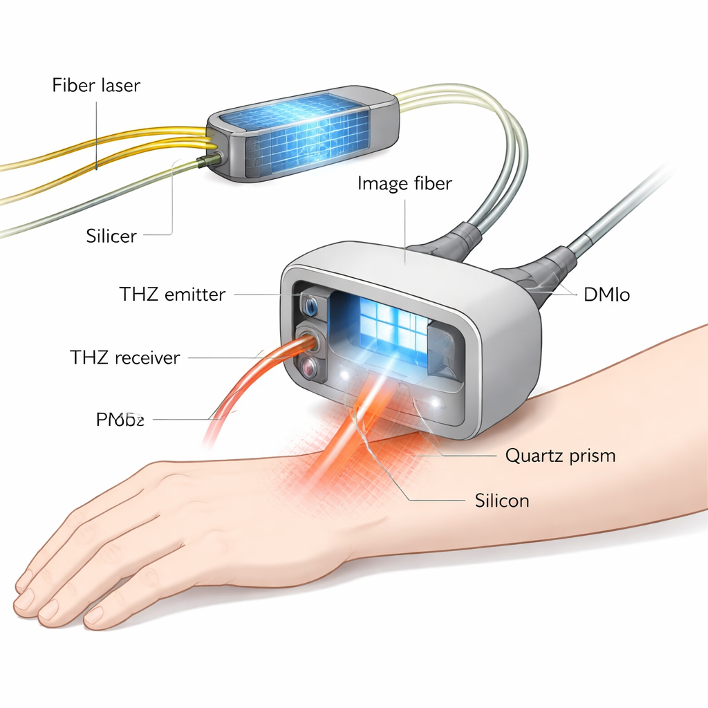
A Compact Probe Driven Entirely by Optical Fibres
The researchers solved these practical barriers by building an all-fibre-coupled terahertz imaging system around a small probe that can be moved to where the patient is. Instead of steering terahertz beams with bulky mirrors in free space, they guide the light that creates and detects the terahertz pulse through flexible optical fibres, similar to those used in telecommunications. Inside the probe, a quartz prism and a thin silicon wafer press against the sample surface. Terahertz waves enter the prism, skim along the silicon–sample interface, and reflect back in a process called attenuated total reflection, which is highly sensitive to the properties of the thin layer of tissue just beneath the probe.
Painting Patterns with Light to Build Images
To avoid slow mechanical scanning, the team uses a “single-pixel” imaging strategy. Rather than measuring each image point separately, they shine a series of carefully designed light patterns onto the silicon wafer using a blue laser and a digital micromirror device, delivered through an imaging fibre bundle. These patterns locally change how the silicon interacts with terahertz waves, effectively imprinting a matching pattern onto the terahertz beam. For each pattern, a single detector records the total reflected terahertz signal, and a computer mathematically reconstructs the image from many such measurements. By choosing patterns based on a special Hadamard matrix and taking advantage of a silicon wafer whose electrical response decays in only a few microseconds, the system can switch patterns at up to 20,000 times per second. This yields video-rate imaging with a spatial resolution of about 360 micrometers—fine enough to resolve small skin features—while achieving more than 30,000 image pixels per second, more than five times faster than previous comparable systems.
Testing on Metal Patterns, Animal Tissue, and Human Skin
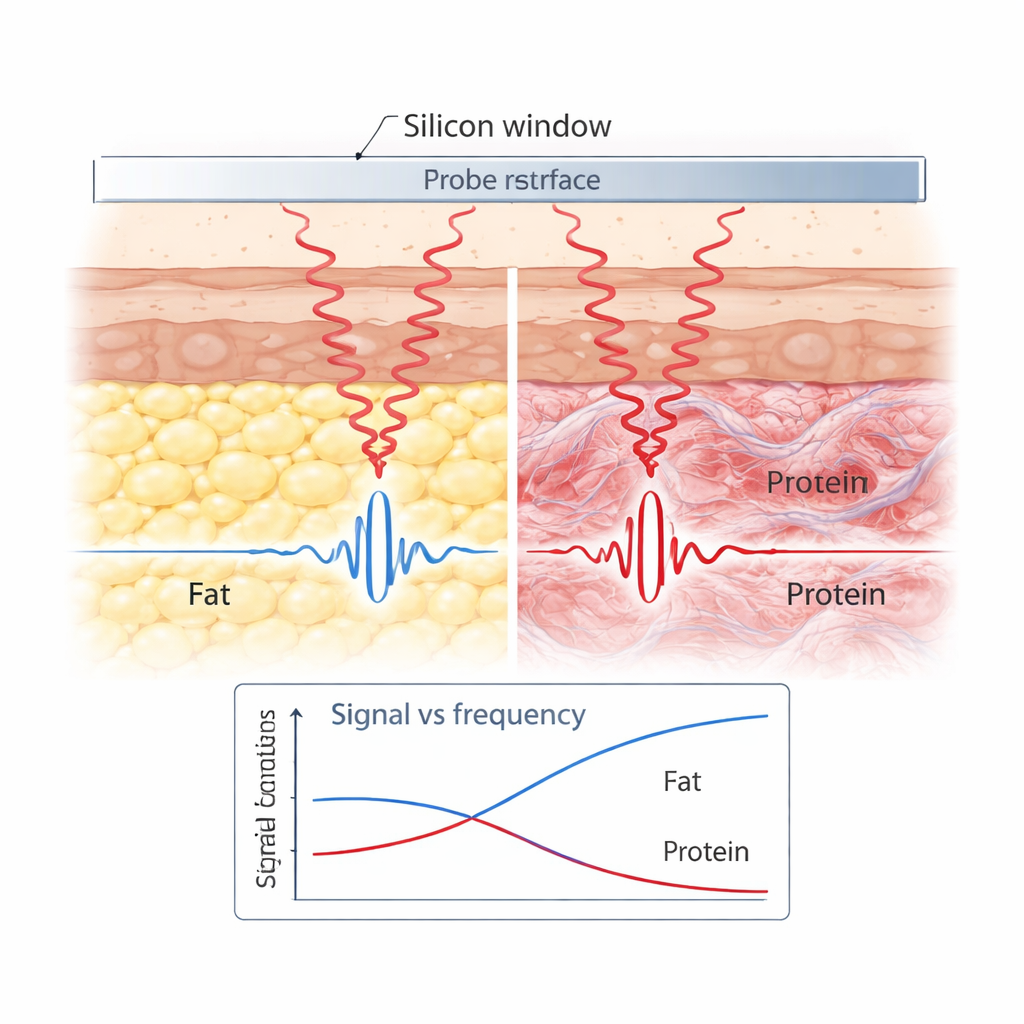
To validate image quality, the authors first imaged a tiny gold “cartwheel” pattern on quartz. The terahertz images clearly showed the metal spokes with high contrast, matching optical photographs and confirming the system’s resolution and stability. Next, they turned to a piece of porcine (pig) tissue containing both fat and protein-rich regions. Because fat holds less water and has different molecular vibrations than protein, the two regions gave distinct terahertz signatures in both signal strength and phase across frequency, allowing a clear boundary between them to be mapped. Finally, the team demonstrated real-time in vivo imaging on a volunteer’s arm. The terahertz probe easily distinguished a dry scab from the surrounding healthy, more hydrated skin, reproducing the scab’s shape and confirming that the technique can work on living tissue in real time.
Faster, Friendlier Scans for Future Clinics
Altogether, this work shows that terahertz imaging can be packaged into a fibre-fed, handheld-style sensor that delivers rapid, non-contact, non-ionizing views of tissue just beneath the skin. By combining attenuated total reflection, single-pixel imaging, and smart use of silicon’s properties, the system achieves high speed, fine detail, and robustness in a compact form factor. With further development, such devices could assist doctors in diagnosing skin cancers, monitoring wound healing, guiding the precise removal of diseased tissue, and even integrating with robotic platforms for automated, safe, and gentle imaging at the bedside.
Citation: Mou, S., Stantchev, R.I., Saxena, S. et al. All-fibre-coupled terahertz single-pixel imaging for biomedical applications. Nat Commun 17, 1571 (2026). https://doi.org/10.1038/s41467-026-68290-x
Keywords: terahertz imaging, single-pixel imaging, biomedical diagnostics, skin cancer, noninvasive spectroscopy