Clear Sky Science · en
Hyperreflective choroidal foci may predict pachychoroid macular atrophy development in central serous chorioretinopathy
Why tiny spots in the eye matter
Central serous chorioretinopathy is a mouthful, but for many working-age adults it means something very simple and frightening: suddenly the center of vision in one eye looks blurry, distorted, or dim. While early cases often clear on their own, some people develop a stubborn, long-lasting form that can quietly erode sight. This study asks a practical question with big implications for patients and doctors alike: can tiny bright spots deep inside the eye act as early warning signs that help predict who will respond to treatment and who risks developing permanent central damage?
How a leaky layer can steal sharp sight
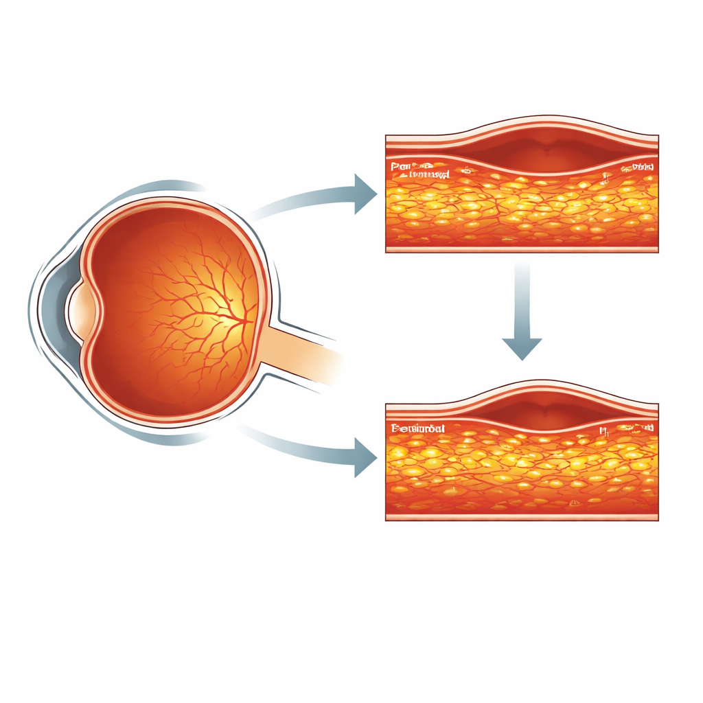
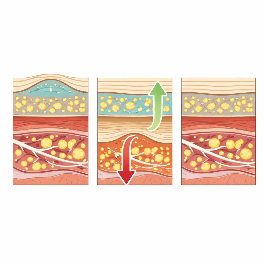
To understand the study, it helps to picture the back of the eye as a layered sandwich. The light-sensing retina sits on top of a rich bed of blood vessels called the choroid, which nourishes it. In central serous chorioretinopathy, this blood-vessel layer leaks fluid under the retina, lifting it up and blurring vision—like a bubble under wallpaper. Modern scanning tools, especially optical coherence tomography (OCT), can slice this region into detailed cross-sections. In some people with this disease, OCT reveals numerous tiny bright specks—“hyperreflective foci”—scattered in the choroid. Doctors suspect these specks mark areas of chronic stress or inflammation. The question is whether counting where they appear, and how they change over time, can tell us how the disease will unfold.
Taking a closer look at problem spots
The researchers reviewed scans from 70 people with long-lasting, previously untreated central serous chorioretinopathy. All had recurrent fluid under the central retina, confirmed by multiple imaging methods. Each person received one of three common treatments: a light-activated drug therapy, an oral medication that affects vessel and fluid balance, or a gentle form of laser. At the start and again after one year, trained graders carefully counted the bright spots in two specific layers of the choroid: an inner zone with medium-sized vessels and a deeper zone with larger channels. They also tracked how much fluid was present, how thick the retina and choroid were, and whether a serious complication—pachychoroid macular atrophy, a form of central thinning and scarring—developed.
When spots shrink versus when they grow
By the end of the year, about half the patients had complete fluid resolution and were labeled responders, while the other half still had persistent or recurring fluid. At the beginning, both groups had similar numbers of bright spots, so a single baseline snapshot could not tell who would respond to treatment. Over time, however, their paths diverged. In responders, the number of spots in both choroidal layers dropped noticeably, in step with better vision, thinner swollen retina, and disappearance of the fluid bubble. In non-responders, spots in the medium-vessel layer actually increased, the choroid failed to thin, and vision tended to worsen. These changes suggest that the spots are not just static scars but dynamic markers of ongoing disease activity: when treatment truly calms the underlying process, the specks fade; when it does not, they accumulate.
Early warning for lasting damage
The most worrying outcome in this illness is macular atrophy—the permanent loss of the central retinal layers that are crucial for reading, recognizing faces, and other fine tasks. None of the patients showed this at the start, but after a year it appeared in just over one in five. It was much more common in those who had not fully responded to treatment. Here the tiny bright spots proved especially telling. People who eventually developed atrophy tended to start with more spots, and by the final visit their total spot count was strongly linked to the presence of damage. In fact, the total number of spots, and especially those in the inner choroidal layer, explained a sizable share of the variation in who developed atrophy. Other measurements—like how thick the choroid was or how tall the fluid bubble rose—were far less informative.
What this means for patients and doctors
For someone living with chronic central serous chorioretinopathy, the study’s message is both cautionary and hopeful. Cautionary, because it shows that even when fluid eventually clears, hidden trouble in the choroid can still set the stage for lasting central damage. Hopeful, because carefully reading those tiny bright specks on standard eye scans can flag higher-risk patients early. Counting how many spots are present at diagnosis, and watching whether they shrink or spread under treatment, could help eye specialists decide who needs closer monitoring, more aggressive therapy, or new treatment approaches. In everyday terms, these microscopic glints in the deep layers of the eye may serve as a traffic light for long-term vision: fewer and fading specks point toward safer ground, while growing clusters warn that the road ahead may lead to permanent loss of sharp sight.
Citation: Pignataro, M.G., Termite, A.C., Borrelli, E. et al. Hyperreflective choroidal foci may predict pachychoroid macular atrophy development in central serous chorioretinopathy. Eye 40, 689–696 (2026). https://doi.org/10.1038/s41433-026-04277-8
Keywords: central serous chorioretinopathy, macular atrophy, choroidal biomarkers, optical coherence tomography, pachychoroid disease