Clear Sky Science · en
Predicting progression to proliferative diabetic retinopathy using automated versus manual quantification of retinal haemorrhages
Why this matters for people with diabetes
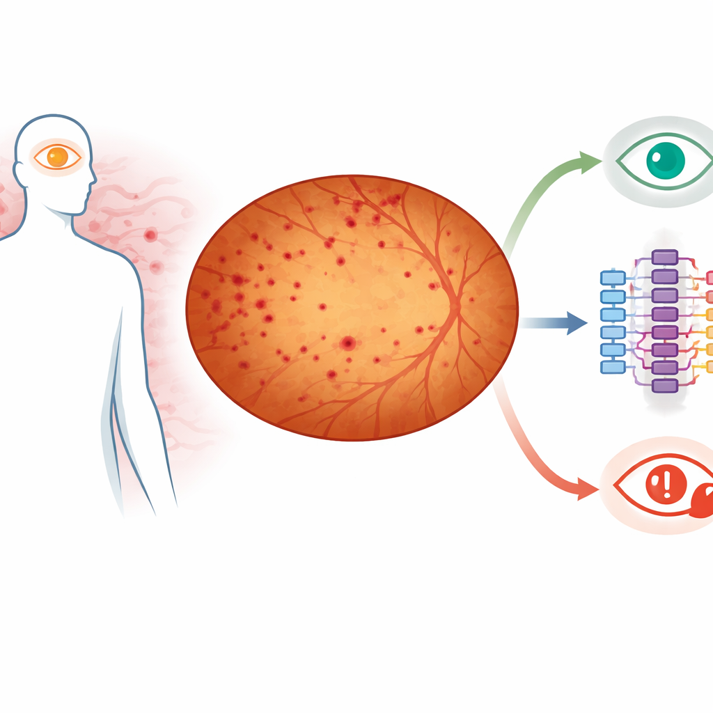
Diabetes can quietly damage the back of the eye long before vision is lost. Doctors know that some people’s eye changes stay mild for years, while others rapidly progress to sight-threatening disease. This study asks a practical question with big implications: can an automated computer system, working from wide-angle photographs of the eye, help predict which patients are most likely to move on to a dangerous stage called proliferative diabetic retinopathy?
Looking deep into the eye
Diabetic retinopathy happens when long-term high blood sugar harms tiny blood vessels in the retina, the light-sensitive layer lining the back of the eye. These vessels can leak or bleed, creating small dark spots called haemorrhages. For decades, eye specialists have judged disease severity by examining a limited central area of the retina in standardized photos. New cameras now capture almost the entire retina in a single ultra-widefield image, revealing many more spots and changes, especially toward the far edges. Earlier work showed that the total area of bleeding and how far those spots lie from the optic nerve—the bright round area where nerve fibers exit the eye—can help predict which eyes are more likely to worsen.
Comparing people versus algorithms
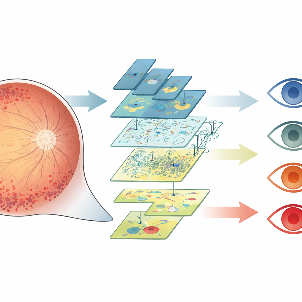
The researchers studied 63 eyes from people with diabetes who had non-proliferative, or not-yet-severe, retinopathy at the start and at least one year of follow-up. Trained experts carefully traced every visible bleeding spot on widefield images using specialized software, a painstaking process that can involve thousands of marks per eye. The same images were then processed by a deep learning–based program called EyeRead, designed to automatically find and outline these spots. For both the human tracings and the automated results, the team calculated how many haemorrhages were present, how much total area they covered, and how far from the optic nerve, on average, the spots were located in both central and peripheral regions.
What the computer saw
The automated system consistently reported fewer haemorrhages and a smaller total area of bleeding than the human graders. This underestimation appeared to stem mainly from how precisely the borders of each spot were drawn, a difficult task because many spots have blurry edges. Still, when the researchers compared eyes one by one, the computer’s measurements and the manual measurements moved together closely, especially for total bleeding area. In other words, even though the absolute numbers were smaller, eyes that humans judged to have more or larger haemorrhages also tended to be scored higher by the algorithm.
Spot location as a warning sign
The most striking finding involved not just how many spots were present, but where they were. Over one year, 29 of the 63 eyes progressed to proliferative diabetic retinopathy, a stage marked by growth of fragile new vessels that can cause serious vision loss. Using statistical models, the researchers found that eyes with haemorrhages located farther from the optic nerve—meaning more toward the retinal periphery—were more likely to progress, regardless of whether the measurements came from human tracers or the automated system. This matches earlier evidence that damage and poor blood flow in the outer retina are a bad sign for future disease.
What this means for future eye care
For people with diabetes and their doctors, the takeaway is encouraging: even an imperfect automated tool, working from wide-angle retinal images, can still help flag eyes at higher risk of advancing to a dangerous stage of disease, especially when it tracks how far bleeding extends toward the outer retina. While the algorithm misses some details compared with expert humans, its measurements line up closely enough to be useful. With further refinement and expansion to other retinal changes, such systems could offer fast, objective risk assessments in busy clinics or remote settings, helping to focus attention and treatment on the patients who need it most.
Citation: Verma, A., Nittala, M.G., Dara, R.M. et al. Predicting progression to proliferative diabetic retinopathy using automated versus manual quantification of retinal haemorrhages. Eye 40, 682–688 (2026). https://doi.org/10.1038/s41433-025-04205-2
Keywords: diabetic retinopathy, retinal imaging, artificial intelligence, disease progression, eye screening