Clear Sky Science · en
Investigation of 3D choroidal components in myopic populations using ultra-widefield OCTA
Why this matters for everyday eyesight
More and more people around the world are becoming nearsighted, especially children and young adults. While glasses or contact lenses can sharpen vision, they do not reveal what is happening deep inside the eye as nearsightedness worsens. This study peeks behind the light‑sensing retina into a hidden, blood‑rich layer called the choroid, using a new wide‑angle scanning technique. By mapping how this layer thins and reshapes as myopia progresses, the researchers hope to explain why some eyes remain stable while others head toward serious, sight‑threatening disease. 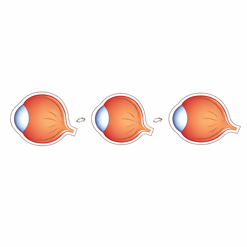
Looking beneath the surface of the eye
The choroid sits behind the retina and is packed with blood vessels and soft supporting tissue. It feeds the outer retina with oxygen and nutrients and helps the eye maintain its shape. In high myopia, this layer is known to thin, and in severe cases abnormal new vessels can grow there, leading to permanent vision loss. Until recently, however, doctors could only examine small central patches of the choroid in detail. The team in this study used an ultra‑widefield version of optical coherence tomography angiography (OCTA), a fast, non‑contact imaging method, to scan a much larger area of the back of the eye and to separate blood‑filled spaces from the surrounding structural tissue in three dimensions.
Who was studied and how the scans were done
The researchers enrolled 147 adults with healthy eyes apart from differing levels of myopia, covering low, moderate, and high ranges of nearsightedness. Both eyes were included when possible. Each participant underwent standard eye tests plus ultra‑widefield OCTA imaging using a high‑speed scanner that captures a 24 by 20 millimeter area of the back of the eye. The choroid was automatically divided into a fine capillary layer near the retina and a deeper layer of larger vessels and supporting tissue. The wide view was then split into a simple three‑by‑three grid covering the central vision area, regions above and below it, and zones closer to the nose and the temple. For each grid square, the team measured how thick the choroid was, how much of its volume was taken up by blood‑filled space, how much by the surrounding tissue, and how dense the smallest vessels were.
How myopia reshapes the deep eye layer
As nearsightedness increased, the choroid became thinner across most regions, especially directly under the center of gaze and in the broader macular area responsible for sharp vision. Total choroidal volume declined in parallel, with the biggest drops again in this central zone. Interestingly, the tiny capillaries in the macular region did not simply fade away. Their flow density actually rose slightly in more myopic eyes, while capillary changes elsewhere were minimal. When the researchers separated blood‑filled volume from the surrounding tissue, they found that the space occupied by larger vessels shrank early, mainly when eyes moved from low to moderate myopia. In contrast, the supporting tissue volume changed more between moderate and high myopia, particularly in and around the macula, hinting that this soft scaffold is lost later in the disease. 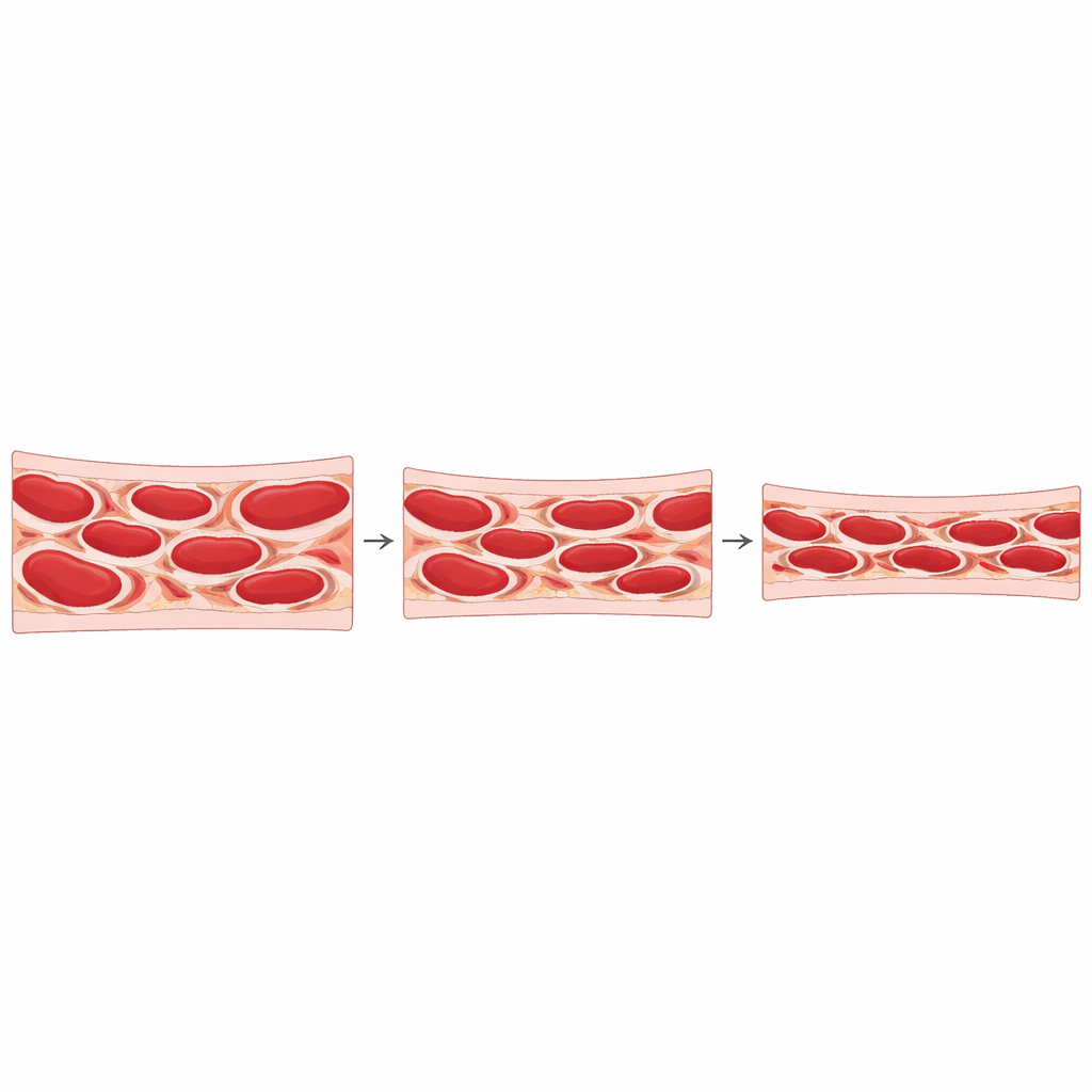
Uneven patterns and what they may mean
The team also examined how eye length and focusing power related to these deep measurements. Longer eyes tended to have thinner choroids and less supporting tissue volume in the central region, while a stronger degree of nearsightedness tracked with multiple choroidal measures. Yet a common index that simply expresses the ratio of vessel volume to total choroidal volume changed only modestly and sometimes even rose as myopia worsened. This happened because both blood‑filled and supporting components shrank together, and in more advanced myopia the supporting tissue appeared to shrink faster. The study revealed that these changes are not uniform: some peripheral regions showed smaller or delayed shifts, suggesting that early myopic remodeling may start in specific areas rather than everywhere at once.
What this means for protecting vision
For a general reader, the takeaway is that myopia is not just about a longer eye or blurrier distance vision. It is also about slow, layered remodeling of the deep blood‑rich coat that nourishes the retina and helps stabilize the eyeball. Using ultra‑widefield OCTA, the researchers showed that central regions crucial for clear sight lose both vessel volume and supporting tissue as myopia progresses, and that the soft scaffold of the choroid particularly thins when eyes cross from moderate into high myopia. Although this study does not yet predict who will develop severe complications, it demonstrates that wide‑angle, three‑dimensional maps of the choroid can reveal early structural warning signs. In the future, such imaging might help doctors track myopic progression more precisely and design treatments that aim not only to correct focus but also to preserve the health of the eye’s hidden support system.
Citation: Rao, T., Yang, J., Liao, Y. et al. Investigation of 3D choroidal components in myopic populations using ultra-widefield OCTA. Eye 40, 630–636 (2026). https://doi.org/10.1038/s41433-025-04203-4
Keywords: myopia, choroid, retinal imaging, OCTA, eye health