Clear Sky Science · en
The axon initial segment-associated microglia regulate neuronal activity and visual perception
Immune Helpers Shaping What We See
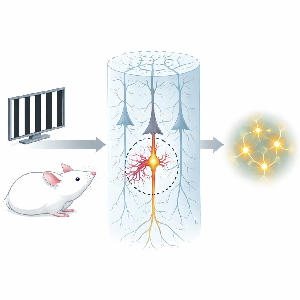
Our ability to see and recognize objects depends on lightning-fast electrical signals in the brain. For many years, most of the credit went to neurons, the classic "wiring" cells. This study reveals that a second, immune-like cell type called microglia quietly helps tune those signals at a critical launch point on neurons. By zooming in on the tiny region where nerve impulses begin, the authors show that a special group of microglia can boost the activity of select neurons and, in doing so, influence how accurately mice tell one visual pattern from another.
Guardians at the Nerve’s “Trigger Zone”
Every excitatory neuron has a short region near its base, the axon initial segment (AIS), where electrical spikes are first generated. The researchers discovered that about one fifth of microglia in the visual cortex form tight, stable contacts with this specific spot, wrapping a process along much of the AIS length. These AIS-associated microglia have distinct shapes and gene-activity profiles compared with other microglia, including higher levels of adhesion and signaling molecules that help them cling to the AIS. One such molecule, integrin β1, appears especially important for forming this snug junction between microglial process and neuron.
How Microglia Give Neurons an Extra Push
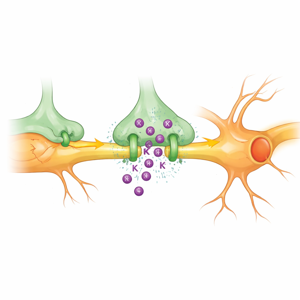
Using paired electrical recordings in brain slices, the team asked whether these "AIS microglia" actually change how their partner neurons fire. Briefly depolarizing a microglial cell that touches the AIS made its associated neuron fire more action potentials in response to the same input, even though there were no synapses between them. This effect did not occur for microglia that touched only the neuron’s cell body or did not touch it at all, pointing to the AIS contact as crucial. Mechanistic experiments showed that when these microglia are depolarized, they release potassium ions through a channel called THIK-1 directly into the tiny gap at the AIS. This small, local rise in potassium causes a subtle depolarization of the neuron’s trigger zone, lowering the input needed to make it fire without disturbing overall synaptic balance.
From Visual Input to Microglial Pulses
To test whether such microglial voltage changes occur naturally, the researchers used fast optical voltage sensors to watch microglia in awake mice viewing drifting visual patterns. Visual stimulation produced brief depolarizing events mainly in microglial processes, not their cell bodies. These events depended on muscarinic receptors, which respond to the neurotransmitter acetylcholine, and on an ion channel called NALCN that lets sodium flow into the microglia. After each depolarization, microglia used THIK-1 to release potassium and restore their resting state. Blocking THIK-1 prevented this recovery, confirming that microglial potassium efflux is a built‑in reset mechanism that is naturally engaged during sensory processing.
Boosting a Small but Powerful Neuronal Subset
Calcium imaging in the visual cortex revealed that only a minority of neurons responded very strongly to moving gratings. These highly responsive cells were often the ones whose AISs were contacted by microglia. When THIK-1 was blocked or removed specifically from microglia, or when microglial depolarization was optically suppressed, the calcium signals of AIS‑associated neurons dropped markedly, while neighboring neurons without AIS contacts were largely unaffected. Disrupting the physical AIS–microglia link by deleting integrin β1 in microglia produced a similar selective loss of strongly responding neurons. In all cases, the overall synchronization and connectivity of whole neuronal ensembles responding to visual stimuli were reduced.
From Cell-to-Cell Contact to Seeing Straight
Finally, the authors asked whether this microscopic partnership matters for behavior. Mice were trained to perform a Go/No-Go visual task, licking for one grating orientation and withholding for another. Once trained, their performance dropped sharply when THIK-1 was blocked in the visual cortex, when THIK-1 was deleted from microglia, or when integrin β1–dependent AIS–microglia contacts were broken. Mice made more false alarms and discriminated orientations less accurately, even though the basic circuitry was intact. These results suggest that a small, specialized set of microglia at the neuron’s trigger zone can selectively amplify key neurons, tighten ensemble coordination, and thereby sharpen visual perception. In essence, immune-derived cells at the AIS act as fine-tuners, using a brief burst of potassium to help the brain decide what it is seeing.
Citation: Wang, Y., Wang, Q., Gao, C. et al. The axon initial segment-associated microglia regulate neuronal activity and visual perception. Cell Res 36, 249–271 (2026). https://doi.org/10.1038/s41422-026-01218-8
Keywords: microglia, axon initial segment, neuronal excitability, visual cortex, potassium signaling