Clear Sky Science · en
Axin1 stabilizes S-opsin and maintains cone photoreceptor survival by inhibiting GSK3β activity
Keeping Our Color Vision Alive
Every time we step into sunlight or glance at a bright screen, tiny cells in our eyes work furiously to turn light into vision. These cells, called photoreceptors, are fragile and can slowly die in many blinding diseases. This study uncovers how a little-known “organizing” protein, Axin1, helps protect cone photoreceptors—the cells that give us daylight and color vision—by keeping key light-sensing molecules in the right place and calming dangerous stress inside the cell.
How Cones See the World
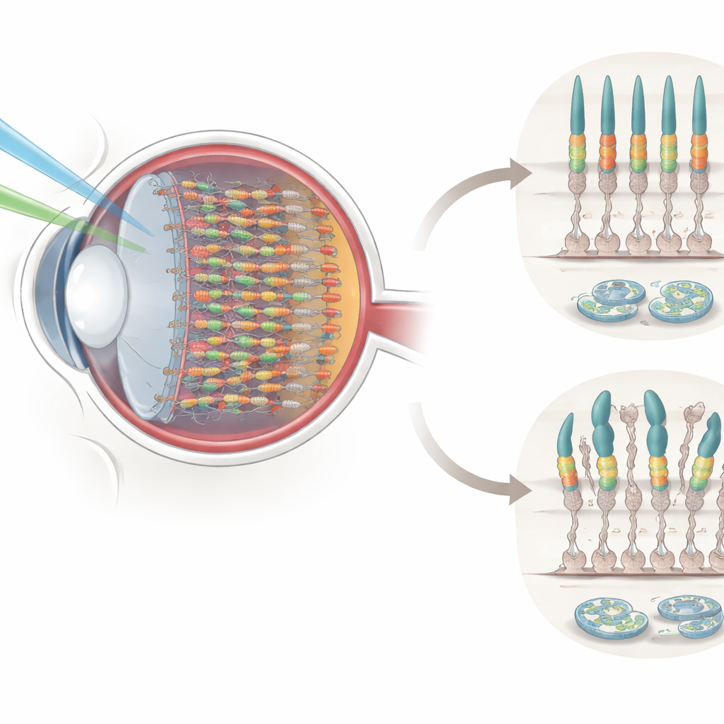
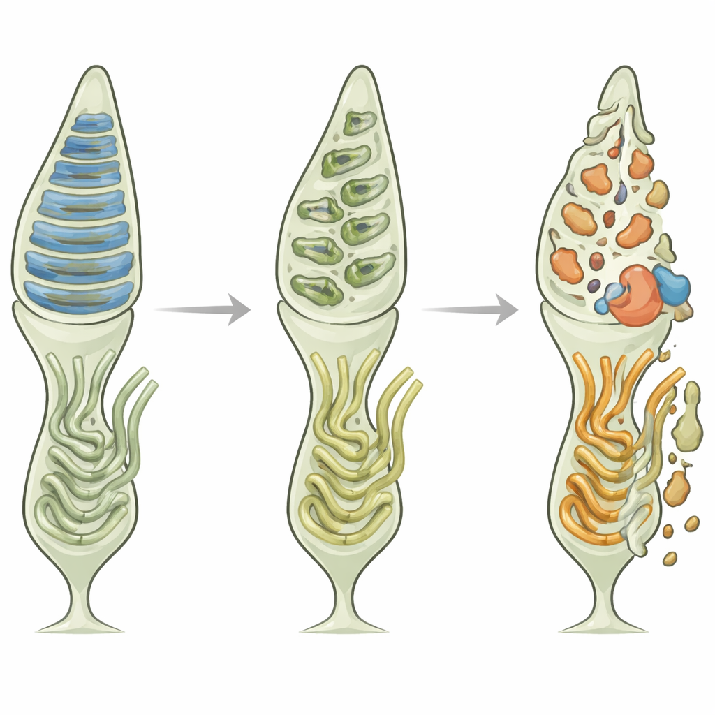
Our retinas contain two main types of photoreceptors: rods for dim, black‑and‑white vision and cones for bright, color vision. Cones carry special pigments, including S‑opsin, which is most sensitive to short‑wavelength (blue) light. These pigments sit in a narrow, stacked region at the tip of each photoreceptor called the outer segment, where incoming light is converted into electrical signals. Because this region is constantly bombarded by light and must renew itself rapidly, it is especially vulnerable to damage and to stress in the cell’s protein‑folding factory, the endoplasmic reticulum (ER). When the ER is overwhelmed by misfolded or misplaced proteins, it can trigger cell suicide, contributing to retinal degeneration and vision loss.
A Hidden Organizer in Cone Cells
The researchers turned their attention to Axin1, a scaffold protein best known for organizing signaling pathways in other parts of the body. Using high‑resolution staining in mouse retinas, they found that Axin1 is not spread evenly across the eye. Instead, it is enriched in cone photoreceptors, especially in the ventral (lower) retina, exactly where S‑opsin is known to be most abundant. Within each cone, Axin1 clusters in the outer segment, closely overlapping with S‑opsin. Over the course of development, Axin1 and S‑opsin appear together, shifting from small spots to elongated rod‑like structures that mark mature cone outer segments. This tight spatial and timing match hinted that Axin1 might be crucial for keeping S‑opsin properly positioned for efficient light detection.
What Happens When Axin1 Is Lost
To test Axin1’s importance, the team selectively removed it from cone cells in mice using a viral gene‑editing approach. These Axin1‑deficient animals showed sluggish pupil constriction when exposed to blue or green light and behaved abnormally in a light–dark preference test, indicating impaired light perception. Under the microscope, their cone outer segments looked disorganized: S‑opsin levels dropped, and instead of forming smooth, rod‑shaped structures, the pigment broke into scattered puncta. The surrounding retinal tissue also showed early signs of trouble. Tight junctions in the supporting pigment layer were disrupted, synaptic markers between cones and downstream neurons were reduced, and glial cells—normally quiet support cells—became activated, all hallmarks of ongoing retinal degeneration.
Stress Inside the Cell and a Dangerous Switch
The absence of Axin1 did not just alter structure; it also intensified biochemical stress. In mouse retinas lacking Axin1, markers of ER stress, such as the protein CHOP, were strongly elevated in photoreceptor layers. In cultured cone‑derived 661W cells, knocking down Axin1 made blue‑light exposure and a chemical ER stressor, tunicamycin, far more toxic, driving up ER‑stress genes and the number of cells undergoing apoptosis. The study linked this vulnerability to a key enzyme, GSK3β, which becomes more active when Axin1 levels fall. Hyperactive GSK3β pushed cells further toward ER stress and death. Conversely, stabilizing Axin1 with a small‑molecule drug, or directly inhibiting GSK3β with lithium chloride, reduced stress markers, calmed GSK3β activity, and rescued many cells from apoptosis.
Turning Protection into Therapy
Taken together, the findings paint Axin1 as a central guardian of cone photoreceptors. By anchoring S‑opsin in the correct place in the outer segment and restraining the stress‑promoting enzyme GSK3β, Axin1 helps cones cope with the relentless demand of bright and short‑wavelength light. When Axin1 is missing or unstable, S‑opsin becomes mislocalized, the ER is flooded with problematic protein, stress pathways flare, and cones are pushed toward degeneration. Because many human blinding disorders ultimately involve loss of cones and ER‑stress‑driven cell death, strategies that boost or mimic Axin1 function—or safely temper GSK3β activity—could offer new avenues to slow or prevent retinal degeneration and preserve our daylight and color vision.
Citation: Xu, J., Man, J., Fan, Y. et al. Axin1 stabilizes S-opsin and maintains cone photoreceptor survival by inhibiting GSK3β activity. Cell Death Discov. 12, 109 (2026). https://doi.org/10.1038/s41420-026-02968-5
Keywords: retinal degeneration, cone photoreceptors, endoplasmic reticulum stress, Axin1, GSK3 beta