Clear Sky Science · en
Comparative phenotypic and molecular profiling of replicative and chemically-induced senescence in articular chondrocytes
Why worn-out joint cells matter
Achy, stiff joints are a common part of getting older, but scientists are discovering that a special kind of “worn-out” cell may be a key driver of osteoarthritis—the most common joint disease worldwide. These cells, called senescent cells, stop dividing and begin to release a cocktail of irritating molecules that can damage nearby tissue. This study asks a deceptively simple but crucial question: when researchers grow cartilage cells in the lab to study osteoarthritis, which methods of making those cells “old” actually resemble what happens in real joints?
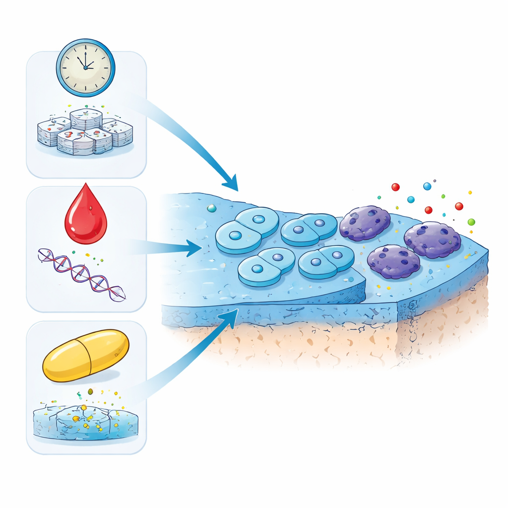
Three different paths to tired cells
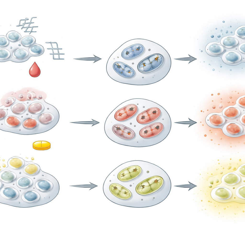
The researchers focused on chondrocytes, the cells that maintain the smooth cartilage coating at the ends of bones. Using cells from sheep joints, they pushed the cells into senescence in three different ways. In one approach, they simply kept the cells dividing over many generations until they reached an old, worn-out state, mimicking aging over time. In the other two approaches, they exposed young cells to low doses of two drugs used in human medicine: doxorubicin, a chemotherapy drug that damages DNA, and dexamethasone, a strong anti-inflammatory steroid injected into painful joints. All three treatments were tuned so that the cells survived long enough to develop a stable, aged-like state.
Common signs of cellular old age
Across all three methods, the chondrocytes displayed classic hallmarks of senescence. They stopped multiplying, showed changes in their cell cycle, and developed a higher activity of a telltale enzyme often used to spot senescent cells. The cells also altered their internal structure, with larger, flatter shapes and changes in the way their DNA is packaged. Deep inside, their mitochondria—the tiny power plants that make cellular fuel—no longer worked efficiently. Using powerful genetic and protein analyses, the team found that key energy-producing pathways and systems for building new proteins were dialed down in every model. Together, these changes paint a consistent picture of cartilage cells that have exited the workforce and are stuck in a metabolically sluggish but persistent state.
Same destination, different routes
Despite these shared features, the three models were far from identical. Cells aged by many rounds of division showed shortened chromosome ends, built up harmful oxygen by-products, lost energy, and released more structural cartilage proteins into their surroundings—features that resemble slow, age-related cartilage wear. In contrast, cells stressed with doxorubicin mounted a strong DNA damage response, activated cell-death machinery, and produced high levels of inflammatory signals, suggesting a harsher, more injury-like scenario. Dexamethasone-treated cells also became senescent but without the same spike in damaging oxygen molecules or apoptosis. Instead, they maintained or even increased their energy output and produced a strong mix of secreted factors linked to inflammation and tissue remodeling, reflecting the complex, double-edged effects of steroid drugs on otherwise healthy cartilage.
Signals sent out into the joint
The substances these senescent cells released—collectively known as the senescence-associated secretory phenotype—varied strongly between models. The long-divided cells secreted fewer classic inflammatory and stress-related proteins but became richer in cartilage matrix components. In contrast, both drug-treated groups, especially the steroid-exposed cells, pumped out many molecules that can attract immune cells, promote inflammation, and reshape the surrounding tissue. These distinct “secretory fingerprints” matter because in a living joint they can either quietly weaken cartilage over decades or drive more aggressive, flare-like damage after injury, chemotherapy, or repeated steroid injections.
What this means for understanding and treating joint disease
To a non-specialist, the key message is that not all aged cartilage cells are created equal. The study shows that while different lab methods can push cartilage cells into an old, senescent state, they do so through different kinds of stress with different consequences. Cells aged by sheer time and repeated division most closely resemble those seen in natural aging and long-standing osteoarthritis. Cells stressed by chemotherapy or steroids better model situations where drugs or acute damage accelerate joint decline. By carefully matching the lab model to the real-world scenario, researchers can better test drugs that clear senescent cells or dial down their harmful secretions. The work also highlights failing mitochondria and disturbed energy balance as a central weakness shared by all senescent cartilage cells—an attractive target for future therapies aimed at keeping our joints healthier for longer.
Citation: Arteaga, M.B., Tarasova, K., Kidtiwong, A. et al. Comparative phenotypic and molecular profiling of replicative and chemically-induced senescence in articular chondrocytes. Cell Death Discov. 12, 106 (2026). https://doi.org/10.1038/s41420-026-02961-y
Keywords: osteoarthritis, cellular senescence, chondrocytes, mitochondrial dysfunction, cartilage degeneration