Clear Sky Science · en
CXCR6+ T cells promote apoptosis and necroptosis in proximal tubules during AKI-to-CKD transition
Why this matters for kidney health
Many people who survive a sudden kidney injury later develop long-term kidney disease, which can eventually lead to dialysis or transplant. Yet doctors still do not fully understand why some kidneys bounce back while others slowly fail. This study uncovers a specific group of immune cells and signals that keep injured kidney tubes from healing, instead pushing them toward scarring and permanent damage. Understanding this hidden drama inside the kidney could open new ways to protect kidney function after severe illness, surgery, or drug toxicity.
From sudden damage to lasting scars
Acute kidney injury (AKI) is a rapid loss of kidney function caused by events such as low blood flow during surgery, severe infection, or toxic drugs. Often, the kidney’s filtering units and their attached tubules can repair themselves. But when the injury is severe or repeated, repair goes wrong. Instead of rebuilding healthy tubules, the tissue shrinks, fills with inflammatory cells, and lays down scar. This shift from short-term injury to chronic kidney disease (CKD) is a major pathway to long-lasting kidney failure, yet the cellular steps that drive it have been unclear.
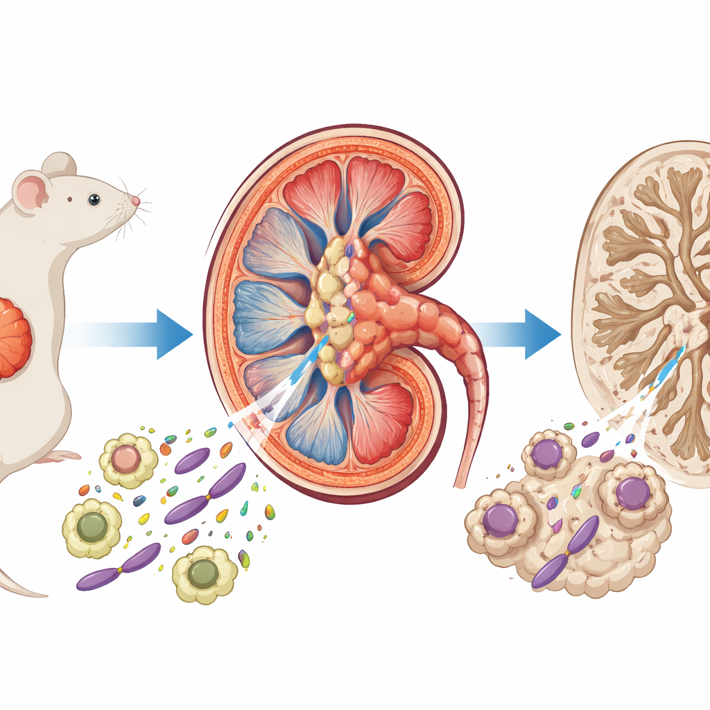
Death signals inside kidney tubules
The authors used a mouse model that mimics poor recovery after kidney injury to examine what happens inside the tiny tubular cells that help reabsorb water and salts. They combined whole-kidney gene profiling, single-cell RNA sequencing, and detailed tissue staining. They found that two forms of programmed cell death—apoptosis, in which cells shrink and quietly break apart, and necroptosis, in which cells swell and rupture—were strongly activated during the weeks after injury. These death signals were especially high in a vulnerable subset of tubular cells that express a surface protein called VCAM-1, which marks them as chronically stressed and prone to atrophy.
Immune cells homing in on injured tissue
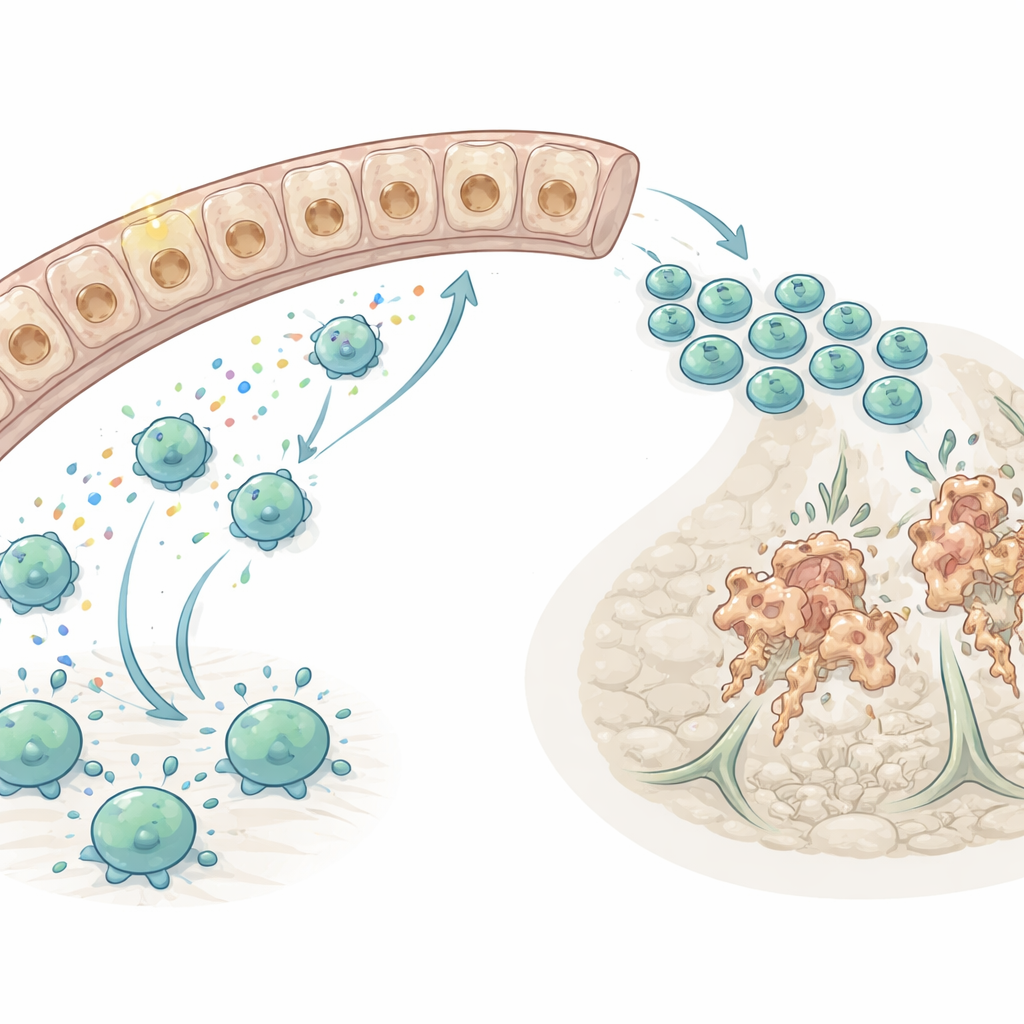
Because immune cells flood into the kidney after injury, the researchers next asked which chemical “homing signals” guide T cells to the damaged tubules. Using computational tools to map cell-to-cell communication from single-cell data, they pinpointed one chemokine pair—CXCL16 (a signal) and CXCR6 (its receptor)—as a dominant pathway drawing T cells into the injured kidney, particularly the CD8 cytotoxic type that can directly kill other cells. They showed that macrophages, a type of tissue-resident immune cell, were the main producers of CXCL16, with injured tubules providing additional signal. In culture, inflammatory messengers such as TNF-α and IL-1β drove macrophages and tubular cells to ramp up CXCL16 through an NF-κB–dependent pathway, linking early inflammation to later T cell recruitment.
A genetic test of the CXCR6 pathway
To test whether this homing route truly worsens injury, the team studied mice lacking CXCR6. Both normal and CXCR6-deficient mice suffered similar initial kidney damage after ischemia, confirming that the early insult was the same. But two weeks later, kidneys without CXCR6 had far fewer T cells, especially killer T cells, around the injured tubules. Markers of cell death—both apoptotic and necroptotic—were substantially reduced, and fewer tubular cells stained positive for DNA fragmentation. At the same time, the damaged kidneys in CXCR6-deficient mice retained healthier tubular markers, showed less cast formation and fibrosis, and had fewer cells stuck in a dedifferentiated, nonfunctional state.
Protecting function, not just structure
Structural improvements matter only if they translate into better kidney performance. To test this, the authors surgically removed the uninjured kidney two weeks after the initial insult, forcing the previously injured kidney to carry the workload. Mice lacking CXCR6 had lower blood urea nitrogen and creatinine levels—standard measures of kidney function—than their normal counterparts, both immediately and over the following days. This shows that blocking CXCR6-bearing T cells not only preserves tubular architecture but also improves the kidney’s ability to filter blood after severe injury.
What this means for future treatments
Overall, the study reveals a harmful feedback loop: inflammation activates macrophages and tubules to release CXCL16, this attracts CXCR6+ T cells, and those T cells intensify tubular cell death and necroinflammatory signaling, promoting scarring and chronic disease. Breaking this CXCL16–CXCR6 axis—or dampening its upstream triggers—could help injured kidneys heal instead of hardening into permanent damage. While these findings come from mice, they highlight a specific immune pathway that may be targeted to slow or prevent the slide from acute kidney injury to chronic kidney disease in people.
Citation: Li, X., Melchinger, I., Chen, Y. et al. CXCR6+ T cells promote apoptosis and necroptosis in proximal tubules during AKI-to-CKD transition. Cell Death Dis 17, 359 (2026). https://doi.org/10.1038/s41419-026-08644-x
Keywords: acute kidney injury, chronic kidney disease, immune cells, tubule cell death, chemokine signaling