Clear Sky Science · en
Radiomics identifies distinct cortical bone texture alterations in patients with CKD using HR-pQCT
Hidden Weakness in Bones
People with chronic kidney disease face a much higher risk of broken bones, yet standard bone scans often say their bones look nearly normal. This study asks a simple but important question: are today’s hospital scanners missing early warning signs of fragile bones in these patients—and can smarter image analysis uncover them before a fracture happens?
Why Kidney Disease Threatens the Skeleton
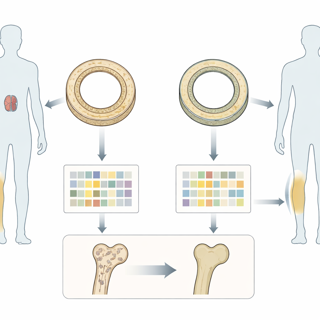
Chronic kidney disease does more than affect blood chemistry; it also quietly reshapes bone. The outer shell of long bones, called cortical bone, normally forms a dense protective ring. In kidney failure, this shell can thin and become more porous and irregular, making bones easier to break. Conventional tools like dual-energy X-ray absorptiometry (DXA) mostly measure overall bone density in 2D and cannot distinguish this outer shell from the spongy interior. Even advanced 3D scans such as high-resolution peripheral quantitative CT (HR-pQCT), which can see tiny pores and thickness, have produced mixed results in clearly separating patients with kidney disease from those without.
A New Way to Read Bone Images
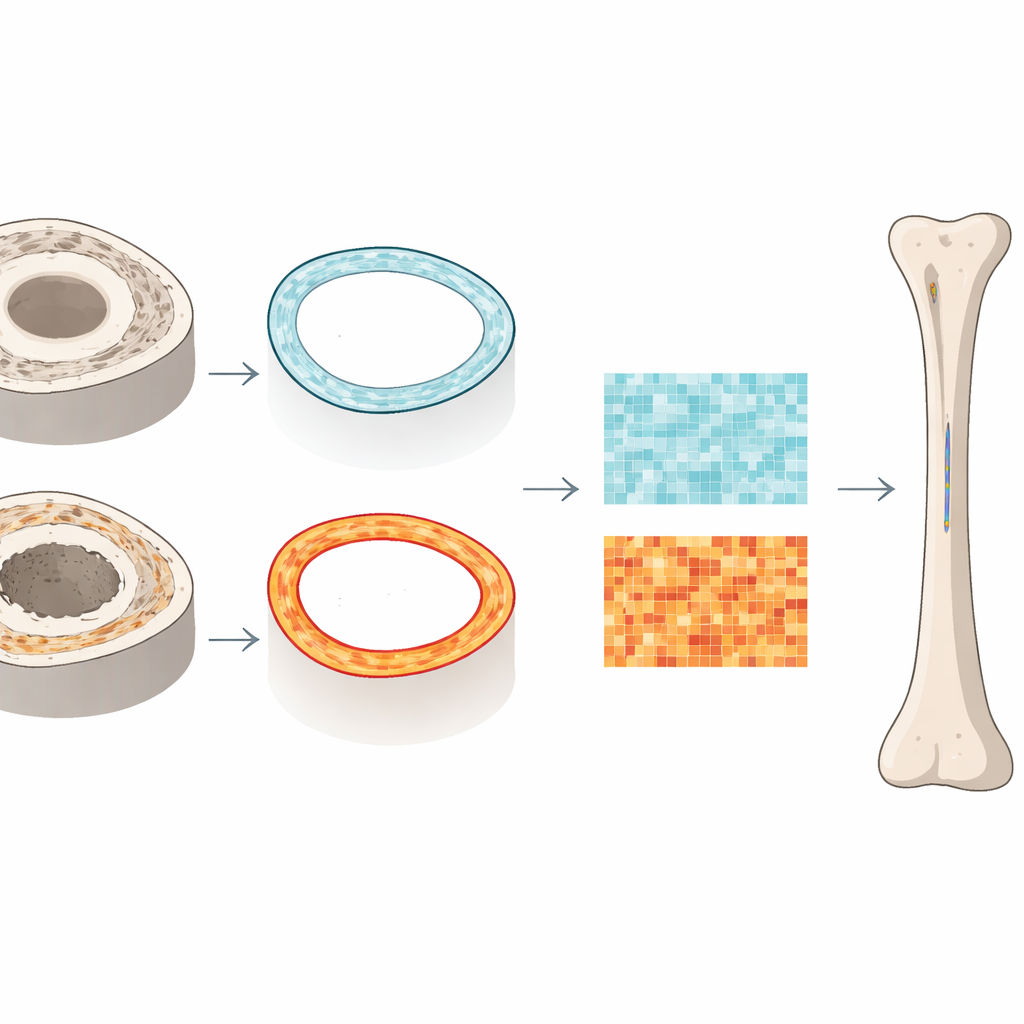
The researchers turned to “radiomics,” a method that treats medical images as rich data maps rather than simple pictures. Instead of only averaging how bright or dark the bone appears, radiomics breaks each scan into hundreds of small mathematical descriptors of patterns, contrast, and texture. These features capture how pixel intensities vary from place to place, revealing subtle irregularities that the eye—or standard software—cannot easily see. Using HR-pQCT scans of the shin bone (tibia) from 72 adults, half with advanced dialysis-dependent kidney disease and half without kidney problems, the team focused specifically on the cortical shell at both the ankle end and the mid-shaft of the bone.
What the Textures Revealed
From more than 24,000 image slices, the radiomics pipeline extracted 753 potential features and then carefully filtered them down to a smaller, non-redundant set. In people without kidney disease, the most informative features were simple ones: basic measures of how bright the bone appeared overall, such as the maximum and minimum gray levels and how much they varied. In those with advanced kidney disease, however, the dominant features came from more complex texture statistics that sense how irregular and patchy the cortex is. Measures related to local “strength” and non-uniformity of neighboring pixels stood out, hinting at a more mottled, disorganized matrix even when standard metrics like density, thickness, and porosity looked quite similar between the two groups.
Zooming In on Subtle Differences
The study also divided each tibia scan into proximal and distal subregions to see whether texture changes clustered in particular areas. In the lower (distal) tibia of kidney disease patients, radiomic measures highlighted regions with stronger local contrast and greater heterogeneity, consistent with increased microscopic pores and disrupted organization. At the mid-shaft (diaphysis), different combinations of features—such as the lowest gray levels and uneven intensity patterns—captured the distinctive signature of kidney-related bone changes. Importantly, these radiomic differences were statistically large and consistent, whereas conventional measurements, including estimates of stiffness and failure load from computer-based mechanical simulations, showed only modest or no group differences.
What This Means for Patients
To a layperson, the key message is that bones in advanced kidney disease can look nearly normal on standard scans while their internal “grain” has already become patchy and weak. By reading HR-pQCT images in a more data-rich way, radiomics detects this hidden roughening of the cortical shell well before it becomes obvious through density loss alone. Although more work is needed in larger and earlier-stage kidney disease populations, this approach could eventually provide doctors with new, noninvasive markers of bone quality—helping to identify patients at high fracture risk sooner and tailor treatments before a serious break occurs.
Citation: Lee, Y., Hong, S., Lee, M. et al. Radiomics identifies distinct cortical bone texture alterations in patients with CKD using HR-pQCT. Bone Res 14, 36 (2026). https://doi.org/10.1038/s41413-026-00515-7
Keywords: chronic kidney disease, bone texture, radiomics, cortical bone, fracture risk