Clear Sky Science · en
Genomic structural variations contribute to inform prognosis in patients with cytogenetically normal acute myeloid leukemia
Why tiny DNA changes matter for leukemia patients
For adults diagnosed with acute myeloid leukemia (AML), doctors increasingly use genetic tests to predict how well treatment is likely to work. Yet for nearly half of these patients, standard chromosome tests look "normal," and their future remains frustratingly hard to forecast. This study shows that hidden, small-scale rearrangements in DNA—too small to be seen under a microscope—can sharply distinguish a group of patients whose disease is far more aggressive than current tools suggest.
Seeing beyond normal-looking chromosomes
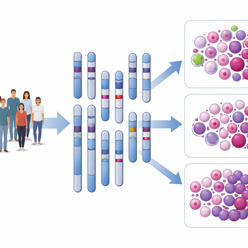
AML is a blood cancer in which immature white blood cells crowd out healthy cells in the bone marrow. Doctors routinely examine chromosomes and known gene mutations to sort patients into favorable, intermediate, or adverse risk groups, which guide choices such as whether to offer a stem cell transplant early. However, about 45% of patients have "cytogenetically normal" AML—meaning their chromosomes appear intact—even though their outcomes range from long-term survival to rapid relapse. The authors reasoned that subvisible DNA rearrangements, known as structural variations, might help explain this hidden diversity.
Using long-read sequencing as a genetic magnifying glass
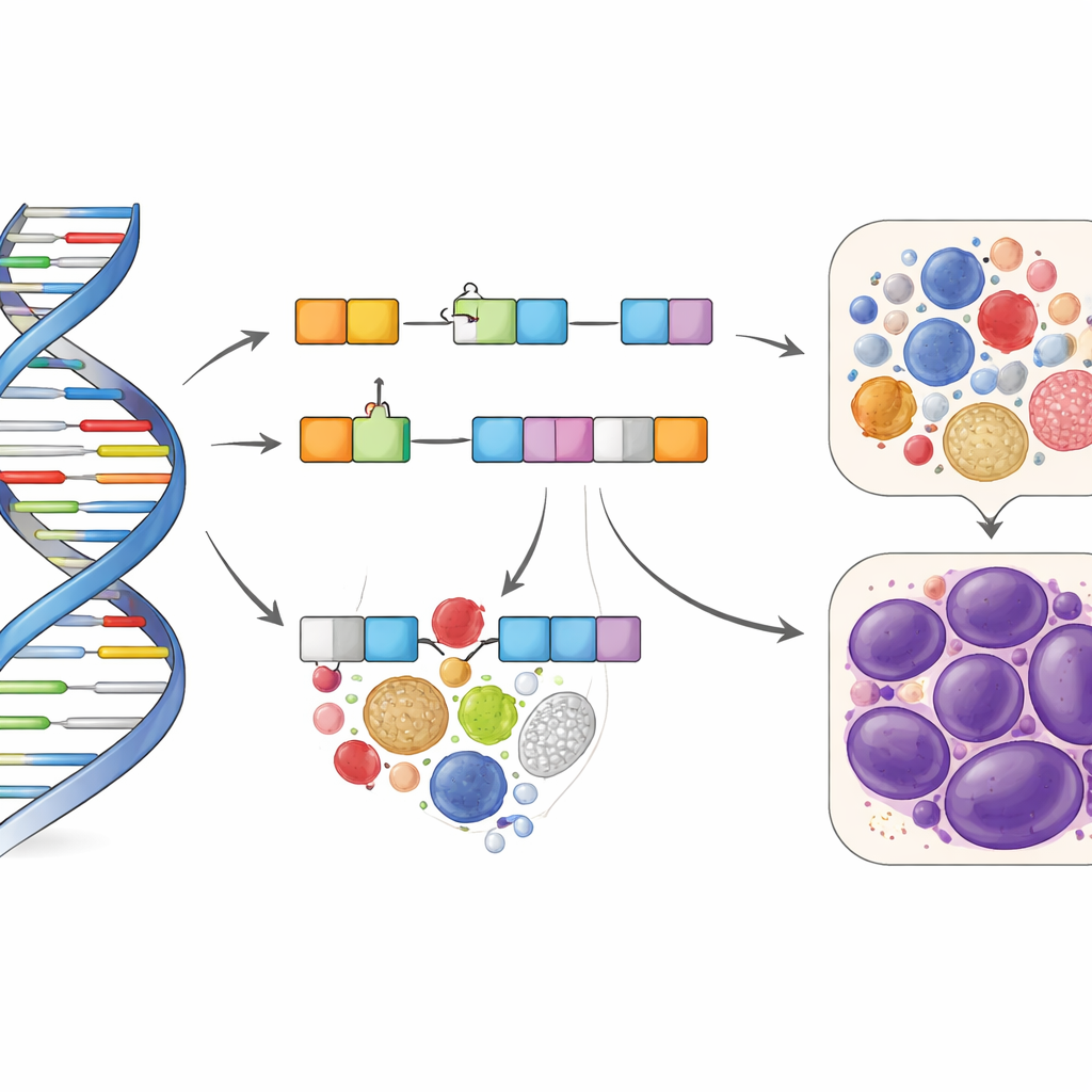
To hunt for these cryptic changes, the team used long-read whole-genome sequencing, a technology that reads very long stretches of DNA and is well suited to spotting insertions, deletions, and other rearrangements. They applied it to leukemia cells from 162 intensively treated adults with cytogenetically normal AML enrolled in two clinical trials. After stringent filtering to remove inherited differences and technical artifacts, they distilled more than two thousand raw findings down to 118 reliable structural variations scattered across the autosomes. Most were small insertions or deletions located in noncoding regions of the genome, such as introns and regulatory zones, rather than directly within protein-coding sequences.
Five small changes define a very high-risk group
The researchers then asked which of these 118 variations tracked with how long patients lived and how long they remained free of events such as relapse or failure to respond to treatment. Using machine learning and survival modeling, they identified five specific structural variations that independently signaled much poorer outcomes. Patients carrying at least one of these "high-risk variations"—about 13–15% of those studied—had lower chances of reaching complete remission, relapsed more often, and had markedly shorter survival, typically under a year. These effects held up even after accounting for well-known mutations such as FLT3 and NPM1 and remained significant in a second, real-world group of 149 additional patients tested with targeted assays.
Improving existing risk scores and explaining why
Current European risk guidelines place all cytogenetically normal patients into broad categories based on certain mutations, but often fail to cleanly separate intermediate from adverse risk. By adding the presence of any of the five high-risk variations as a new "very adverse" tier, the authors created an updated four-level scheme. This refined score predicted overall and event-free survival more accurately than the standard model, especially for patients with NPM1 mutations, who are usually labeled favorable but did very poorly when they also carried these structural changes. Laboratory studies supported a biological basis: the high-risk variations were linked to altered activity of nearby genes, and experimentally changing the levels of some of these genes in cell models disrupted normal cell growth and cell-cycle control, features compatible with more aggressive leukemia behavior.
What this means for patients and care
In everyday terms, this work suggests that some AML patients whose chromosomes look normal and who currently receive an "intermediate" or even "favorable" label may, in fact, harbor a much more dangerous form of the disease. A small set of tiny DNA rearrangements—detectable with modern sequencing or focused follow-up tests—can identify these patients early. Recognizing this hidden very high-risk group could help doctors steer them toward more intensive or experimental treatments and closer monitoring, with the goal of improving outcomes that are currently dismal.
Citation: Bartalucci, N., Mannelli, F., Tarantino, D. et al. Genomic structural variations contribute to inform prognosis in patients with cytogenetically normal acute myeloid leukemia. Blood Cancer J. 16, 37 (2026). https://doi.org/10.1038/s41408-026-01465-3
Keywords: acute myeloid leukemia, structural variation, genomic risk stratification, long-read sequencing, NPM1 mutation