Clear Sky Science · en
Magnetic resonance texture alterations in the caudate nucleus following 18 weeks of clozapine treatment in patients with treatment-resistant schizophrenia
Why this brain study matters for everyday lives
Many people with schizophrenia find relief with standard medications, but a large minority do not. For them, the drug clozapine is often the last and best option—yet doctors still do not fully understand how it changes the brain, or why some patients improve while others do not. This study uses advanced MRI image analysis to look deep inside a small brain region called the caudate nucleus, aiming to spot subtle tissue changes that ordinary scans miss. By doing so, the researchers hope to uncover how clozapine reshapes the brain and why its benefits vary from person to person.
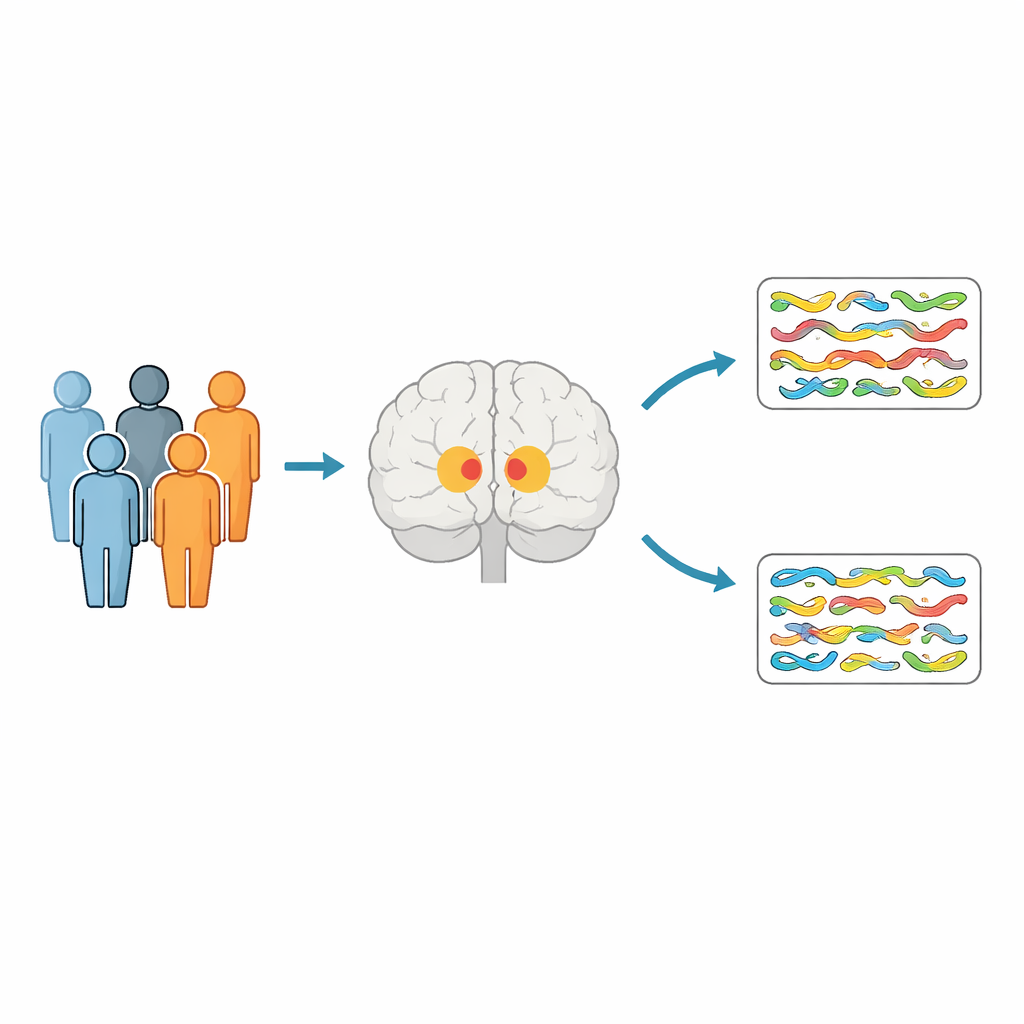
Two groups of patients, one powerful medicine
The researchers followed 64 adults with schizophrenia over 18 weeks. One group had treatment-resistant schizophrenia: their symptoms had not improved despite trying at least two standard antipsychotic drugs. These patients were started on clozapine, and later divided into two subgroups—those who improved on clozapine and those who did not. The second group consisted of people whose symptoms were already well controlled on regular antipsychotic medications and who stayed on the same doses throughout the study. All participants had detailed MRI brain scans at the start and again after 18 weeks, along with repeated symptom ratings.
Looking beyond brain size to brain texture
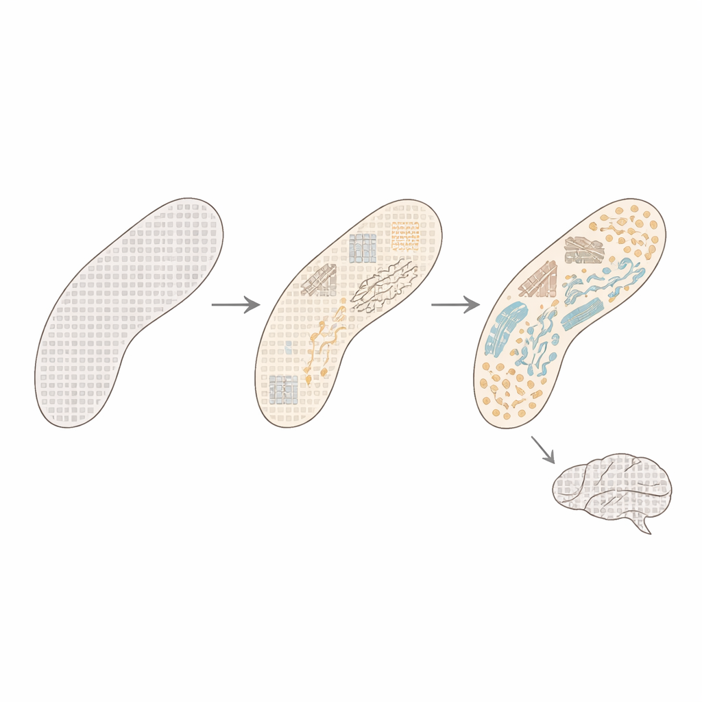
Most earlier imaging studies focused on the size or volume of brain regions, which can miss finer-scale changes in the tissue itself. This work instead applied a method called texture analysis to the caudate nucleus. In simple terms, the method examines how MRI signal intensities vary from one tiny voxel (3D pixel) to its neighbors, capturing patterns of smoothness or complexity that can reflect the micro-organization of cells, fibers, and connections. The team concentrated on a particular texture measure, called correlation, which indicates how similar neighboring voxels are: high values suggest a more uniform tissue pattern, while lower values point to a more varied, complex microstructure.
Clozapine reshapes the caudate’s micro-patterns
After 18 weeks, patients on clozapine showed clear changes in the texture of the left caudate nucleus, whereas the control group on stable standard medication did not. In both clozapine responders and non-responders, the correlation measure in the left caudate decreased over time compared with the control group. This pattern implies that clozapine exposure, regardless of whether symptoms improved, was linked to a shift toward a more heterogeneous and complex tissue pattern in this key brain region. Importantly, these changes were not seen in the cerebellum, a comparison region that is less affected by antipsychotic drugs, suggesting the effect was specific to the caudate.
Hints of hidden brain differences within resistant illness
Even before clozapine treatment began, the subgroup that later responded to clozapine showed higher caudate texture correlation than both the non-responders and the control patients. That is, their caudate tissue looked more uniform at baseline. Over 18 weeks, both clozapine subgroups moved toward more complex patterns, but the ways these baseline features related to symptom change differed. In responders, a more uniform caudate at the start was tied to less improvement in certain positive symptoms. In non-responders, a more uniform caudate was instead linked to better overall and general symptom improvement. These contrasting links suggest that, beneath the shared label of treatment-resistant schizophrenia, there may be distinct underlying brain types that shape how people respond to clozapine.
What this means for understanding and treating schizophrenia
This study shows that clozapine can alter the fine-grained structure of the caudate nucleus within just a few months, in ways that standard MRI measures of brain size may not detect. The observed shift toward more complex tissue patterns may reflect subtle remodeling of nerve cell branches, connections, and myelin, or protection against damage from chemical imbalances such as excess glutamate. While the exact biology still needs confirmation from future work, texture analysis emerges here as a promising tool for tracking how powerful psychiatric medications reshape the brain. In the long run, such sensitive MRI markers could help clarify why some patients benefit from clozapine while others do not—and move psychiatry closer to tailoring treatment to each person’s unique brain profile.
Citation: Jo, W., Moon, S.Y., Sim, H. et al. Magnetic resonance texture alterations in the caudate nucleus following 18 weeks of clozapine treatment in patients with treatment-resistant schizophrenia. Transl Psychiatry 16, 203 (2026). https://doi.org/10.1038/s41398-026-03967-x
Keywords: treatment-resistant schizophrenia, clozapine, caudate nucleus, brain MRI, texture analysis